Comparative Study
. 2010 Jan 1;79(1):57-66.
doi: 10.1016/j.bcp.2009.07.016. Epub 2009 Aug 4.

Role of cytochrome P450 2E1 in protein nitration and ubiquitin-mediated degradation during acetaminophen toxicity

Affiliations
Comparative Study

Role of cytochrome P450 2E1 in protein nitration and ubiquitin-mediated degradation during acetaminophen toxicity

Mohamed A Abdelmegeed et al. Biochem Pharmacol. .

Abstract

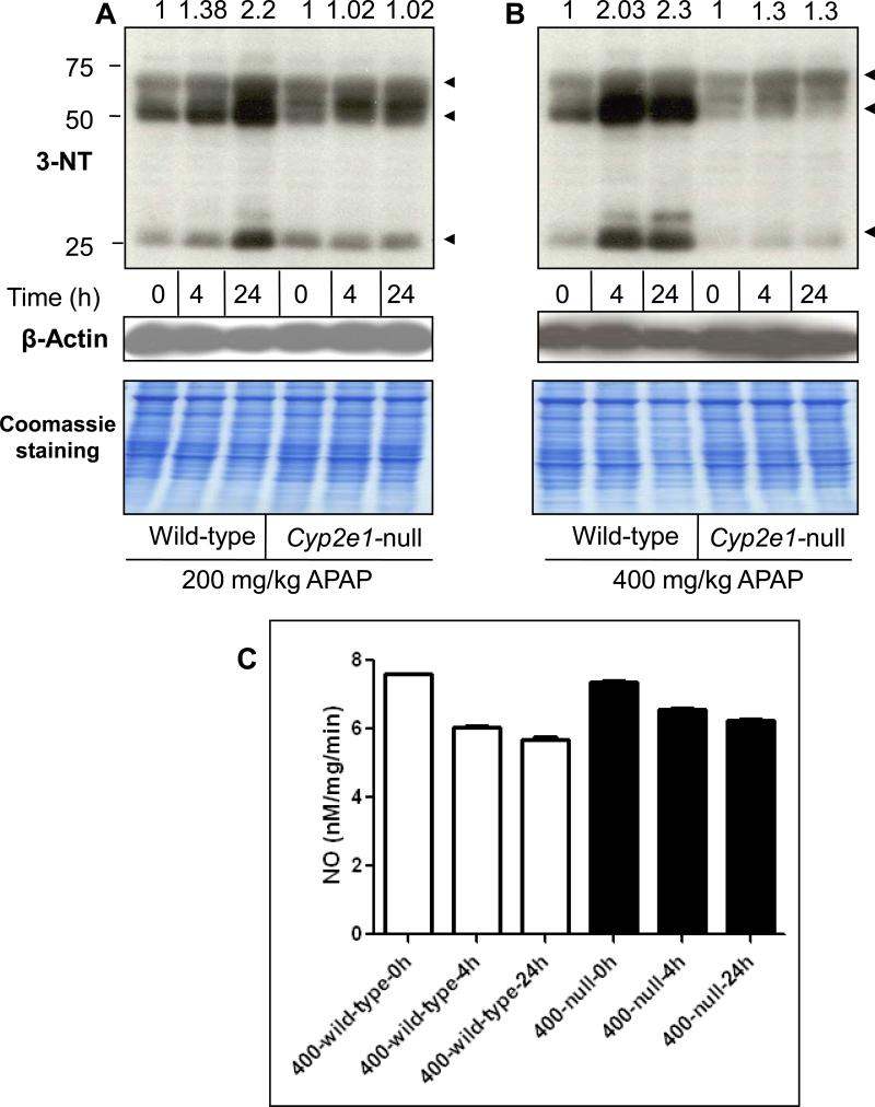
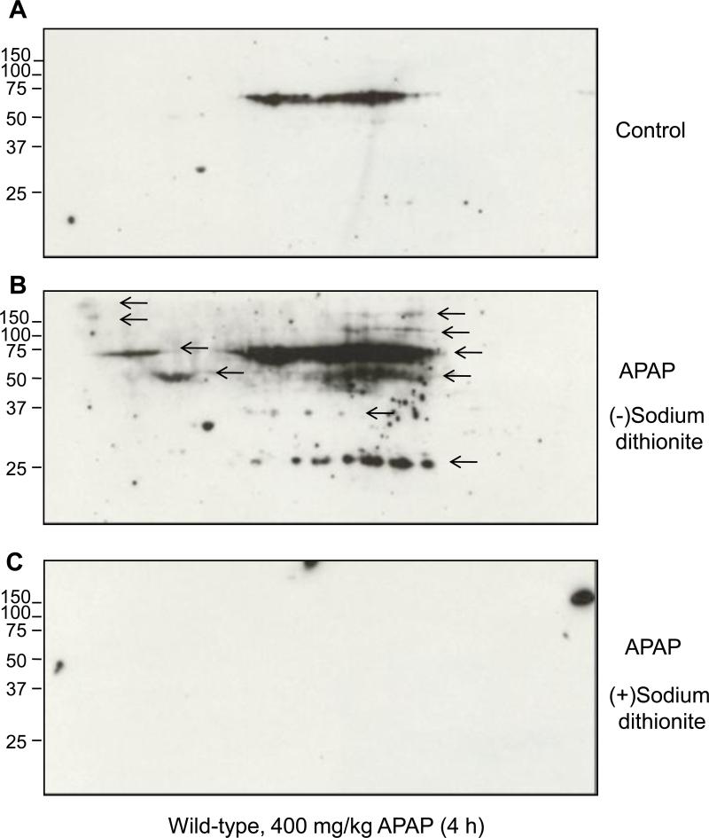
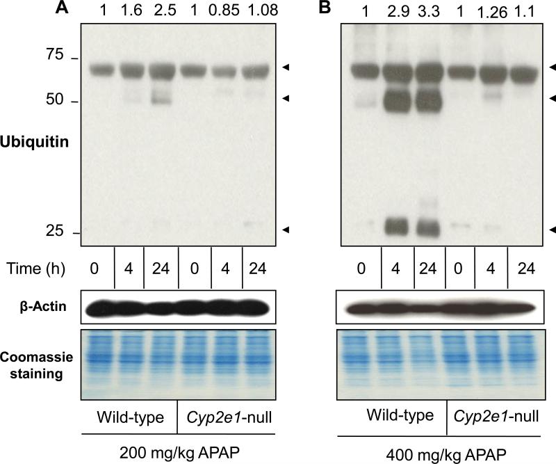
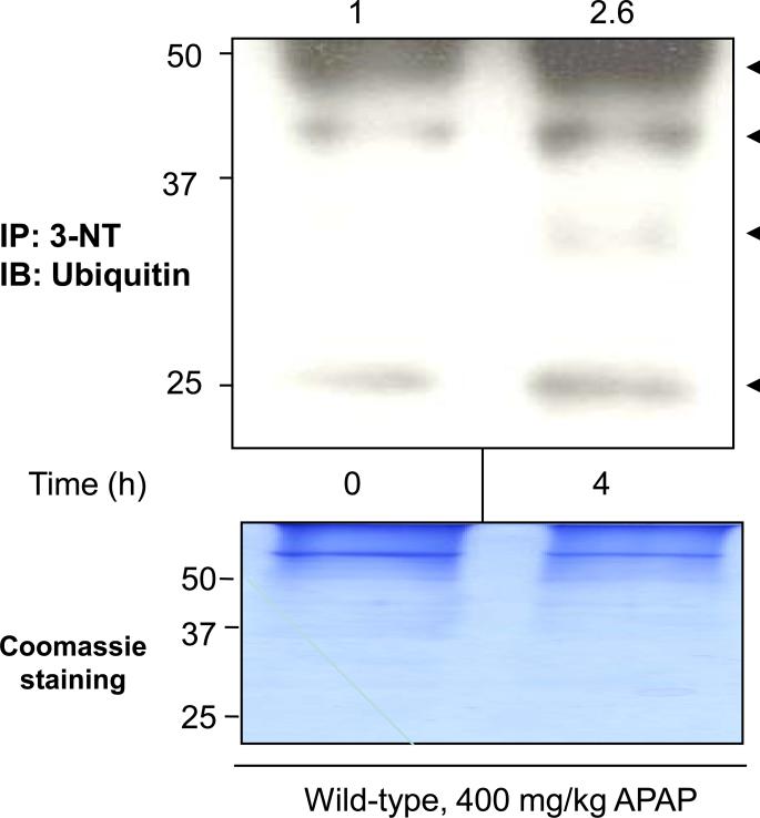
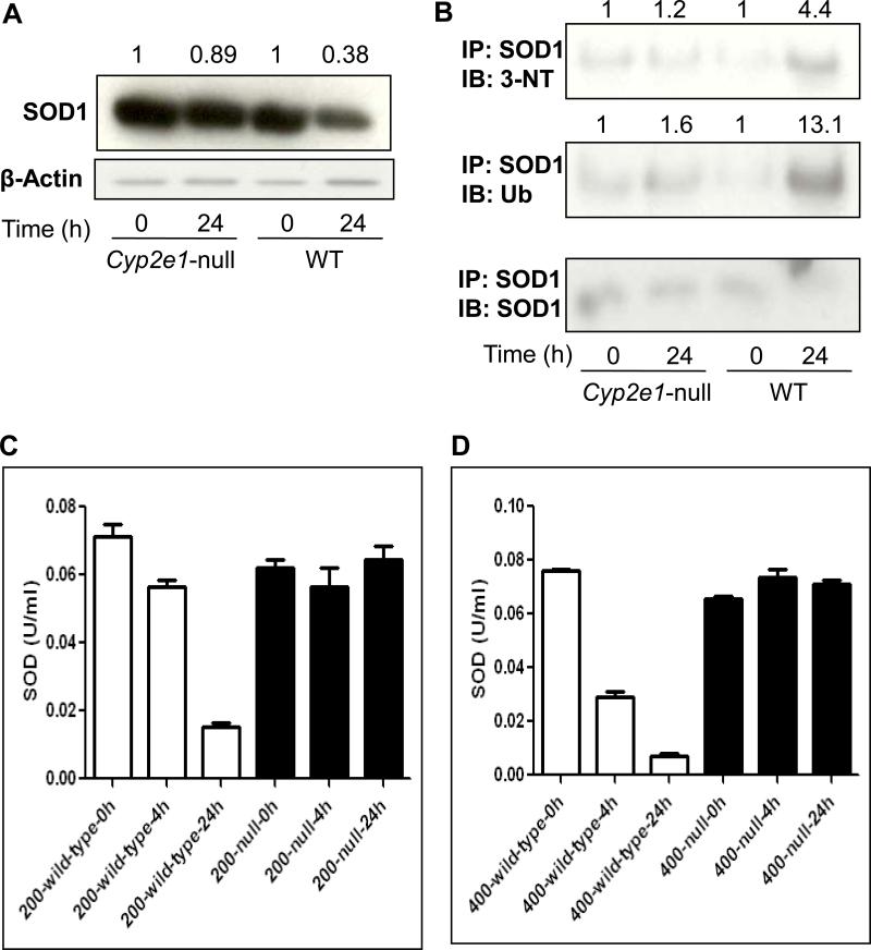
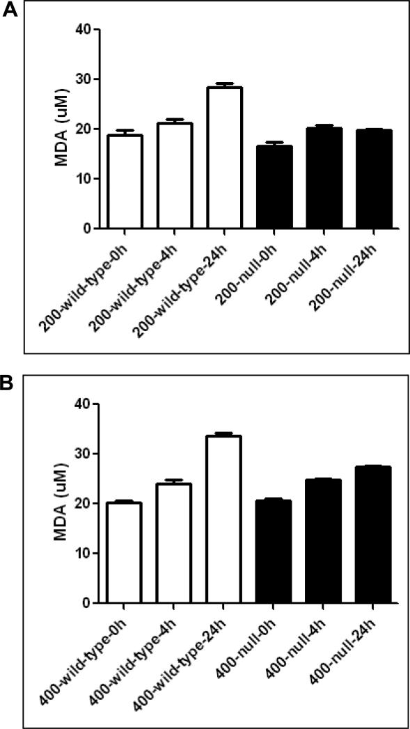
It is well established that following a toxic dose of acetaminophen (APAP), nitrotyrosine protein adducts (3-NT), a hallmark of peroxynitrite production, were colocalized with necrotic hepatic centrilobular regions where cytochrome P450 2E1 (CYP2E1) is highly expressed, suggesting that 3-NT formation may be essential in APAP-mediated toxicity. This study was aimed at investigating the relationship between CYP2E1 and nitration (3-NT formation) followed by ubiquitin-mediated degradation of proteins in wild-type and Cyp2e1-null mice exposed to APAP (200 and 400mg/kg) for 4 and 24h. Markedly increased centrilobular liver necrosis and 3-NT formation were only observed in APAP-exposed wild-type mice in a dose- and time-dependent manner, confirming an important role for CYP2E1 in APAP biotransformation and toxicity. However, the pattern of 3-NT protein adducts, not accompanied by concurrent activation of nitric oxide synthase (NOS), was similar to that of protein ubiquitination. Immunoblot analysis further revealed that immunoprecipitated nitrated proteins were ubiquitinated in APAP-exposed wild-type mice, confirming the fact that nitrated proteins are more susceptible than the native proteins for ubiquitin-dependent degradation, resulting in shorter half-lives. For instance, cytosolic superoxide dismutase (SOD1) levels were clearly decreased and immunoprecipitated SOD1 was nitrated and ubiquitinated, likely leading to its accelerated degradation in APAP-exposed wild-type mice. These data suggest that CYP2E1 appears to play a key role in 3-NT formation, protein degradation, and liver damage, which is independent of NOS, and that decreased levels of many proteins in the wild-type mice (compared with Cyp2e1-null mice) likely contribute to APAP-related toxicity.


Figures

Fig. 1
Fig. 1. Effect of APAP on hepatic necrosis in wild-type and Cyp2e1-null mice
Photomicrographs (200×) after H&E staining from the left hepatic lobes from the indicated mouse livers are presented. Liver histology was performed for: wild-type mice (A-C) or Cyp2e1-null mice (D-F) exposed to 200 mg/kg APAP for 0, 4, and 24 h, respectively. Liver histology was also performed for wild-type mice (G-I) or Cyp2e1-null mice (J-L) exposed to 400 mg/kg APAP for 0, 4, and 24 h, respectively. PC, pericentral regions; PP, periportal regions. Arrows indicate severe necrosis in the PC regions.
Fig. 2
Fig. 2. Levels of hepatic protein nitration and NOS activity in wild-type and Cyp2e1-null mice following APAP treatment
Equal amounts of liver cytosolic proteins (40 μg/well) from different groups were separated on 12% SDS–PAGE, transferred to nitrocellulose membranes, and subjected to immunoblot analysis by using anti-3-NT antibody (A and B, upper panels) and anti-β-actin antibody (A and B, middle panels) for normalization. Coomassie-stained gels are also presented (A and B, lower panels) to further demonstrate equal protein loading. The relative ratio of the 3-NT level detected between the APAP-exposed samples and the corresponding control, which was set at 1, is shown in the top. Equal amounts of liver cytosolic proteins were used to measure total NOS activity (C) according to the manufacturer's instructions.
Fig. 3
Fig. 3. 2D-PAGE and immunoblot analysis for 3-nitrotyrosine detection in hepatic proteins
Immunoreactive nitrated proteins in cytosolic proteins (300 μg/gel) are shown from untreated wild-type mice (A) or treated (B) with 400 mg/kg APAP for 4 h. Immunoblot results (A and B) are before reduction, while (C) represents the result after reduction in the presence of sodium dithionite.
Fig. 4
Fig. 4. Levels of hepatic protein ubiquitination in wild-type and Cyp2e1-null mice following APAP exposure
Equal amounts of liver cytosolic proteins (40 μg/well) from different groups were separated on 12% SDS–PAGE, transferred to nitrocellulose membranes, and subjected to immunoblot analysis by using anti-ubiquitin (A and B, upper panels) and anti-β-actin antibody (A and B, middle panels) for normalization. Coomassie-stained gels are also presented (A and B, lower panels) to further demonstrate equal protein loading. The relative ratio of the 3-NT level detected between the APAP-exposed samples and the corresponding control, which was set at 1, is shown in the top.
Fig. 5
Fig. 5. Ubiquitin conjugation of nitrated proteins in APAP-exposed wild-type mice
Equal amounts of cytosolic proteins (1 mg/sample) in saline control (left lane) and wild-type mice treated with 400 mg/kg APAP for 4 h (right lane) were immunoprecipitated with the monoclonal antibody against 3-NT. The immunoprecipitated proteins were then subjected to immunoblot analysis using the specific antibody against ubiquitin (arrows in top panel). A Coomassie-stained gel is also presented to demonstrate equal protein loading (bottom panel). The relative ratio of the ubiquitin immunoreactivity detected between the control (set as 1) and APAP-exposed samples is shown in the top.
Fig. 6
Fig. 6. Levels of cytosolic SOD1, nitrated SOD1, ubiquitinated SOD1, and activity in wild-type and Cyp2e1-null mice treated with APAP
Equal amounts of or cytosolic proteins (20 μg protein/well) from different groups were separated on 15% SDS–PAGE, transferred to nitrocellulose membrane, and subjected to immunoblot analysis by using the specific anti-SOD1 antibody (A, upper panel) or β-actin (A, lower panel). Density of SOD1 band in each lane was calculated by quantitative densitometry, normalized to that of the corresponding β-actin bands. The relative ratio is presented for the SOD1 level detected in the corresponding control group, which was set at 1, and APAP-exposed groups. Immunoprecipitated proteins from each group were then subjected to immunoblot analysis with the anti-3-NT (B, top panel), anti-ubiquitin (Ub) (B, middle panel), or anti-SOD1 antibody (B, bottom panel). Density of 3-NT or ubiquitin bands was normalized to that of the corresponding SOD1 band. The relative ratio of the immunoreactivity against each target protein detected between the control (set as 1) and APAP-exposed samples is shown in the top. The enzyme activity of SOD was measured using commercially available kit following the manufacturer's protocol in both wild-type and Cyp2e1-null mice treated for 4 or 24 h with 200 mg/kg (C) or 400 mg/kg APAP (D).
Fig. 7
Fig. 7. Levels of hepatic MDA in wild-type and Cyp2e1-null mice subjected to treatment with APAP
Equal amounts of liver cytosolic proteins (0.5 mg/sample) were used to measure the levels of MDA according to the manufacturer's protocols.

Similar articles

Cited by

References

    1. Prescott LF. Hepatotoxicity of mild analgesics. Br J Clin Pharmacol. 1980;10(Suppl 2):373S–9S. - PMC - PubMed
    1. Thomas SH. Paracetamol (acetaminophen) poisoning. Pharmacol Ther. 1993;60:91–120. - PubMed
    1. Ostapowicz G, Fontana RJ, Schiodt FV, Larson A, Davern TJ, Han SH, et al. Results of a prospective study of acute liver failure at 17 tertiary care centers in the United States. Ann Intern Med. 2002;137:947–54. - PubMed
    1. James LP, Mayeux PR, Hinson JA. Acetaminophen-induced hepatotoxicity. Drug Metab Dispos. 2003;31:1499–506. - PubMed
    1. Kon K, Kim JS, Jaeschke H, Lemasters JJ. Mitochondrial permeability transition in acetaminophen-induced necrosis and apoptosis of cultured mouse hepatocytes. Hepatology. 2004;40:1170–9. - PubMed

Publication types