. 2009 Nov;220(1):57-70.
doi: 10.1016/j.expneurol.2009.07.023. Epub 2009 Aug 3.

High frequency stimulation can block axonal conduction

Affiliations

High frequency stimulation can block axonal conduction

Alicia L Jensen et al. Exp Neurol. 2009 Nov.

Abstract

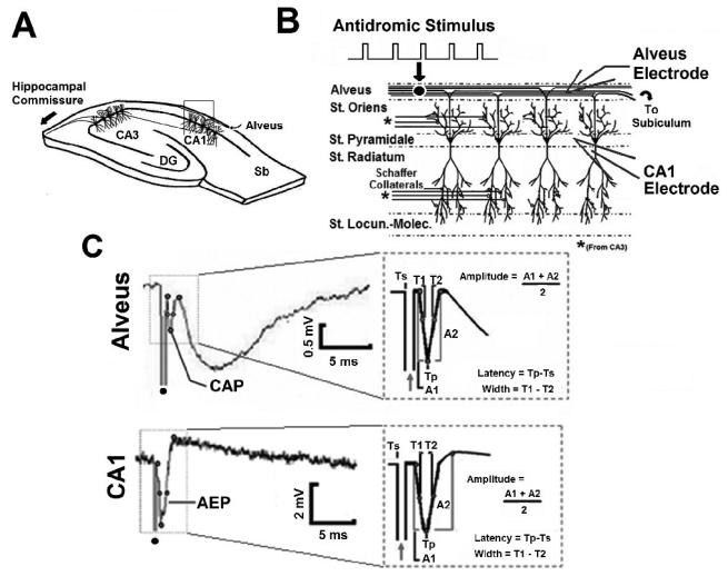
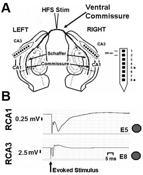
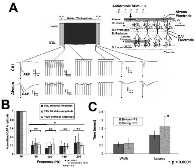
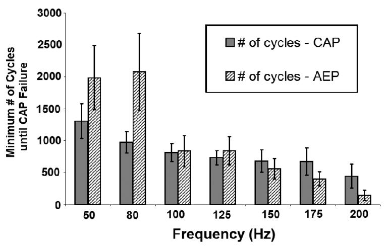
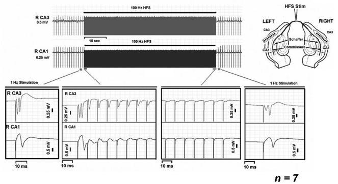
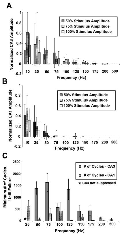
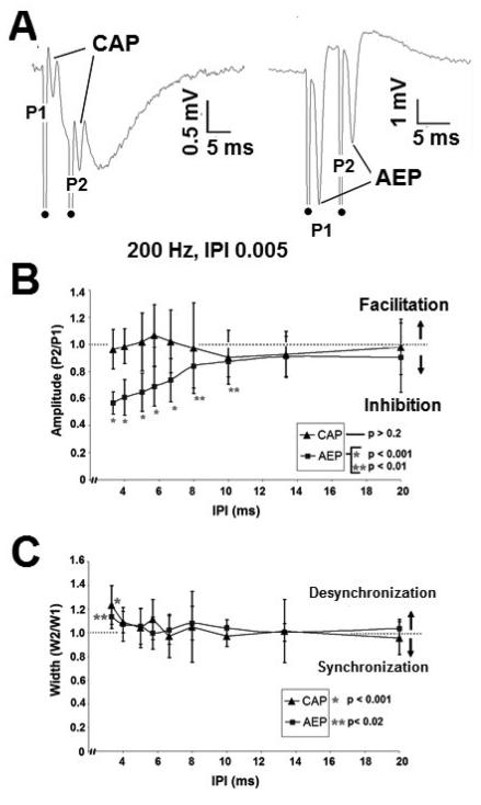
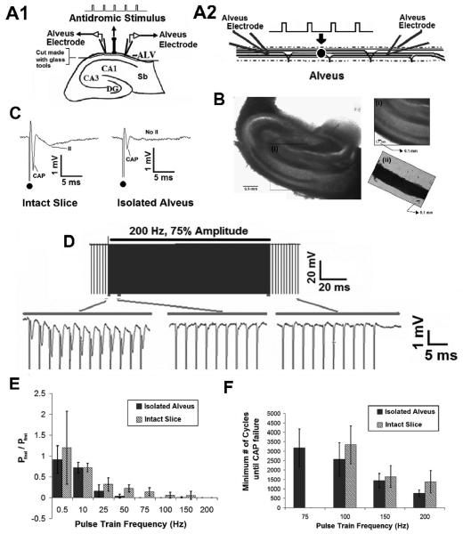
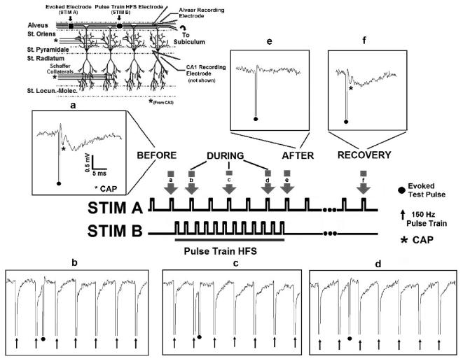
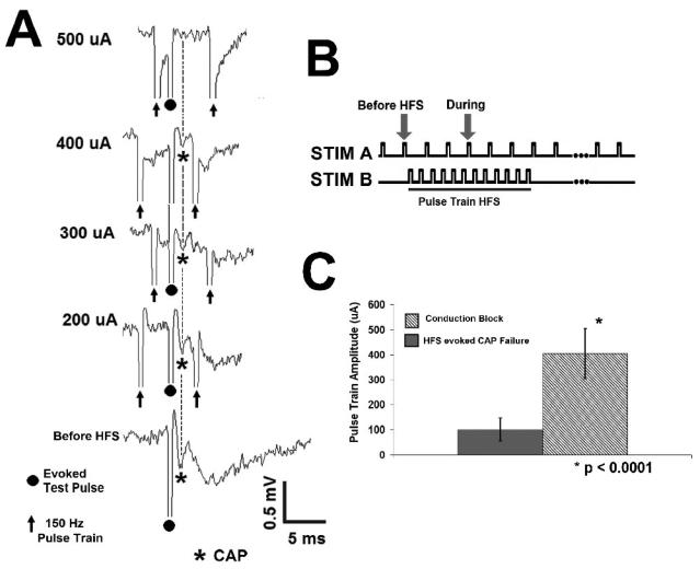
High frequency stimulation (HFS) is used to control abnormal neuronal activity associated with movement, seizure, and psychiatric disorders. Yet, the mechanisms of its therapeutic action are not known. Although experimental results have shown that HFS suppresses somatic activity, other data has suggested that HFS could generate excitation of axons. Moreover it is unclear what effect the stimulation has on tissue surrounding the stimulation electrode. Electrophysiological and computational modeling literature suggests that HFS can drive axons at the stimulus frequency. Therefore, we tested the hypothesis that unlike cell bodies, axons are driven by pulse train HFS. This hypothesis was tested in fibers of the hippocampus both in-vivo and in-vitro. Our results indicate that although electrical stimulation could activate and drive axons at low frequencies (0.5-25 Hz), as the stimulus frequency increased, electrical stimulation failed to continuously excite axonal activity. Fiber tracts were unable to follow extracellular pulse trains above 50 Hz in-vitro and above 125 Hz in-vivo. The number of cycles required for failure was frequency dependent but independent of stimulus amplitude. A novel in-vitro preparation was developed, in which, the alveus was isolated from the remainder of the hippocampus slice. The isolated fiber tract was unable to follow pulse trains above 75 Hz. Reversible conduction block occurred at much higher stimulus amplitudes, with pulse train HFS (>150 Hz) preventing propagation through the site of stimulation. This study shows that pulse train HFS affects axonal activity by: (1) disrupting HFS evoked excitation leading to partial conduction block of activity through the site of HFS; and (2) generating complete conduction block of secondary evoked activity, as HFS amplitude is increased. These results are relevant for the interpretation of the effects of HFS for the control of abnormal neural activity such as epilepsy and Parkinson's disease.


Figures

Fig. 1
Fig. 1. Field potentials evoked by hippocampal fiber tract stimulation in-vitro
A. In-vitro experimental schematic. Sb – Subiculum, DG- Dentate Gyrus. B. Extracellular field potentials were recorded simultaneously in the CA1 stratum pyramidale (CA1 electrode) and alveus (Alvear Electrode). C. Simultaneously recorded compound action potential (CAP) and antidromic evoked potential (AEP) in-vitro in response to antidromic stimulation of the alveus. Dot denotes stimulus artifact. Inset: AEP and CAP amplitude, width, and latency measurements.
Fig. 2
Fig. 2. Field potentials evoked by commissural fiber tract stimulation in-vivo
A. Hippocampal potentials (CA1, CA3) were recorded bilaterally in response to commissural fiber stimulation in-vivo. B. Field potentials generated in response to commissural tract stimulation from right hippocampus are shown. Top trace, evoked field response from CA1. Bottom trace, evoked field response from CA3. Dot denotes stimulus artifact.
Fig. 3
Fig. 3. Evoked axonal responses fail to follow HFS pulse trains in-vitro
A. Evoked responses (CAP) failed to follow prolonged pulse train HFS. The frequency of the evoked stimulus given before and after HFS was 0.5 Hz. The example illustrates the effect of a 200 Hz pulse train. HFS generates initial excitation that decreases in amplitude over the stimulus duration, until the evoked response is gone. Dot denotes stimulus artifact during 0.5 Hz control stimulation. B. Failure of HFS evoked excitation is frequency dependent (p<0.001, ANOVA), with responses unable to faithfully follow pulse train HFS above 50 Hz. Effects on somatic activity (AEP) were similar (not shown). C. CAP width and latency increased prior to complete failure (mean +/− SD; p<0.016, p<0.0001, respectively, student's paired t test), AEP width and latency were unchanged (p<0.2, student's paired t test; not shown).
Fig. 4
Fig. 4. Minimum number of pulse train cycles until CAP failure
The minimum number of cycles until CAP failure decreases as HFS frequency increases, and was dependent on stimulus frequency but not stimulus amplitude (p<0.0001, p>0.5, respectively, ANOVA).
Fig. 5
Fig. 5. Pulse train HFS of fiber tracts in-vivo
The antidromic (CA3) and orthodromic (CA1) evoked responses failed to follow pulse train HFS of the commissural fiber tract (100 Hz). The frequency of the evoked stimulus given before and after HFS was 0.5 Hz. Inset, schematic of the in-vivo experimental arrangement.
Fig. 6
Fig. 6. Failure to follow pulse trains in-vivo is frequency and amplitude dependent
A, For antidromic activity (CA3), evoked activity decreased as frequency of stimulation was increased (p<0.0001, ANOVA, as compared across all stimulation frequencies), while stimulus amplitude had a slightly significant effect (p<0.017, CA3; ANOVA, as compared across all frequencies of stimulation). B. For orthodromic activity (CA1), evoked activity decreased as frequency of stimulation was increased (p<0.0001, ANOVA, as compared across all stimulation frequencies), while stimulus amplitude had a slightly significant effect (p<0.031, CA1; ANOVA, as compared across all frequencies of stimulation). C. The minimum number of pulse train cycles until complete failure was dependent on stimulus frequency for antidromic activation (CA3) (p<0.001, as compared across stimulation frequencies, ANOVA), but not orthodromic activation (p>0.1, as compared across stimulation frequencies, ANOVA).
Fig. 7
Fig. 7. Paired pulse paradigms in-vitro
A. Paired pulse responses generated by 200 Hz (IPI 5 ms) applied to the alveus. Responses recorded extracellularly in the alveus (left) and CA1 pyramidal layer (right). Dot denotes stimulus artifact. B. Cellular field responses (AEP) demonstrated paired pulse inhibition above 100 Hz (IPI 10 ms), while alvear field responses (CAP) were unaffected by the paired pulse paradigm. Paired pulse amplitude ratio, (P2/P1). C. CAP and AEP width were unaffected by the paired pulse paradigm up to IPIs of 4 ms. Below an IPI of 4 ms, cellular (AEP) and axonal (CAP) paired pulse responses shown slight desynchronization. Paired pulse width ratio, (W2/W1).
Fig. 8
Fig. 8. Evoked Axonal Responses are unable to follow HFS in the Isolated Alveus
A1 Transverse hippocampal slice. A2. Schematic of the isolated alvear network showing stimulating and recording electrode placement. B. Photograph of transverse hippocampal slice. (i) Inset: Magnification of the alvear axon field in the intact slice. (ii). Inset: Isolated alvear axon field. C. Example of the evoked alvear potential in the intact slice and isolated alveus. In the isolated preparation, the CAP is present but the secondary synaptic activation (II) is abolished, in response to an evoked stimulus. D. Evoked axonal activity (CAP) were unable to follow HFS even after isolation of the alvear axon field in-vitro (200Hz, 75% Amplitude). E. Failure to follow pulse train stimulation was frequency dependent. Changes in amplitude where analyzed by normalizing the amplitude of the final pulse in the train to the first pulse in the train (n=5). F. No statistically significant difference in the number of cycles until CAP failure between the intact slice and isolated alveus was observed using pulse trains above 75 Hz. In both cases, the number of cycles until CAP failure decreased as stimulus frequency increased (p>0.9, ANOVA, comparing isolated alveus and full slice).
Fig. 9
Fig. 9. Pulse train HFS blocks axonal conduction in-vitro
Evoked test pulses were generated by a stimulating electrode placed in the alvear axon field. An independent electrode, located in the alveus, applied the pulse train HFS. Field potentials were recorded extracellularly in the alveus and CA1 somatic layer (see inset describing hippocampal network). (a) A robust evoked response (CAP, denoted by asterisk) was generated by STIM A (0.5 Hz) before HFS. Pulse train HFS (STIM B, 500 μA, 150Hz) blocks axonal conduction. Insets, b through d (evoked response, STIM A,). (e, f) Evoked responses (CAP) generated by STIM A are depressed following termination of HFS, but recover over ~ 60 seconds following HFS termination (n=8).
Fig. 10
Fig. 10. Pulse train HFS Conduction Block is Amplitude Dependent
A. Conduction block is amplitude dependent, with complete block occurring at higher stimulus amplitudes. B. Stimulation paradigm in-vitro. C. The threshold current for complete block is significantly larger than the stimulus amplitude at which evoked responses fail to follow pulse train HFS (405 ± 101 μA, 101 ± 46 μA, respectively, p<0.0001, Student's 2 sample t test).

References

    1. Abelson J, Curtis G, Sagher O, Albucher R, Harrigan M, Taylor S, Martis B, Giordani B. Deep brain stimulation for refractory obsessive-compulsive disorder. Biological Psychiatry. 2005;57:510–516. - PubMed
    1. Anderson TR, Hu B, Iremonger K, Kiss ZHT. Selective Attenuation of Afferent Synaptic Transmission as a Mechanism of Thalamic Deep Brain Stimulation-Induced Tremor Arrest. J Neurosci. 2006;26:841–850. - PMC - PubMed
    1. Bar-Gad I, Elias S, Vaadia E, Bergman H. Complex Locking Rather Than Complete Cessation of Neuronal Activity in the Globus Pallidus of a 1-Methyl-4-Phenyl-1,2,3,6-Tetrahydropyridine-Treated Primate in Response to Pallidal Microstimulation. J Neurosci. 2004;24:7410–7419. - PMC - PubMed
    1. Bellinger S, Miyazawa A, Steinmetz P. Submyelin potassium accumulation may functionally block subsets of local axons during deep brain stimulation: a modeling study. J Neural Engineering. 2008;5:263–274. - PubMed
    1. Benabid A, Benazzouz A, Hoffmann D, Limousin P, Krack P, Pollak P. Long-term electrical inhibition of deep brain targets in movement disorders. Movement Disorders. 1998;13:119–125. - PubMed

Publication types

MeSH terms