Comparative Study
. 2009 Nov;36(2):320-30.
doi: 10.1016/j.nbd.2009.07.023. Epub 2009 Aug 4.

Mice deficient in dihydrolipoyl succinyl transferase show increased vulnerability to mitochondrial toxins

Affiliations
Comparative Study

Mice deficient in dihydrolipoyl succinyl transferase show increased vulnerability to mitochondrial toxins

Lichuan Yang et al. Neurobiol Dis. 2009 Nov.

Abstract

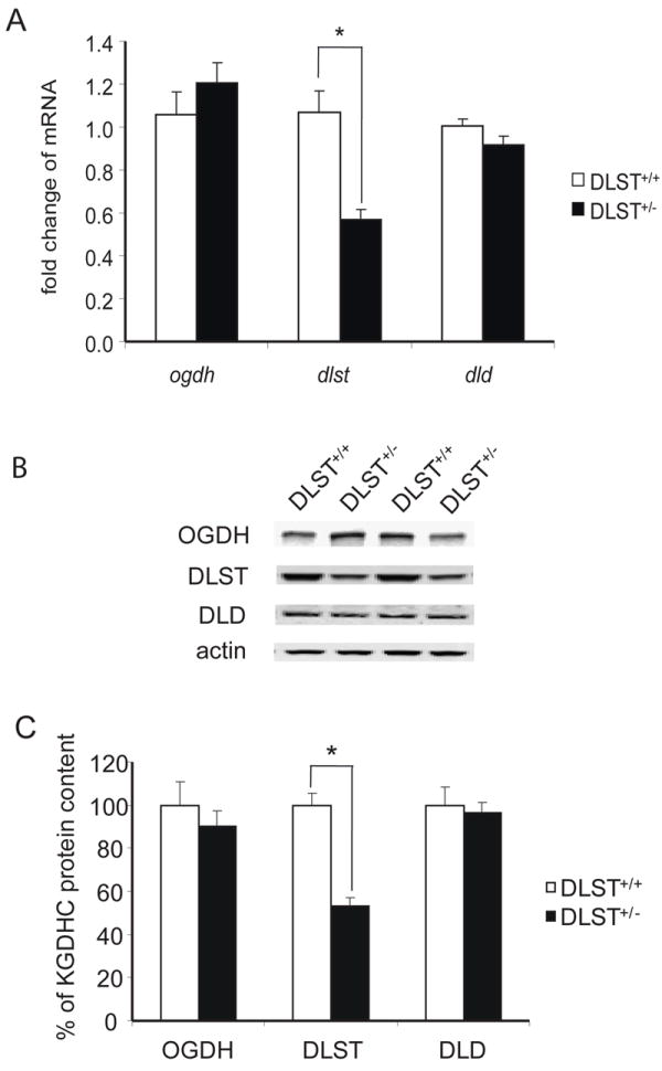
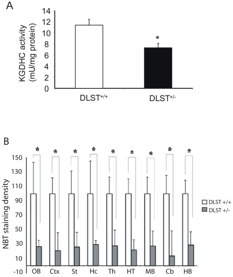
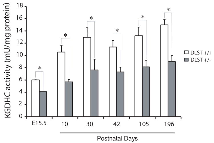
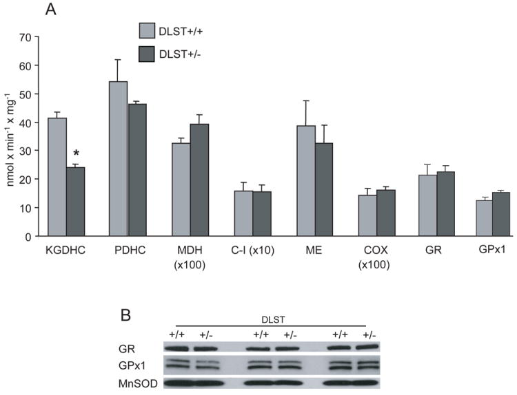
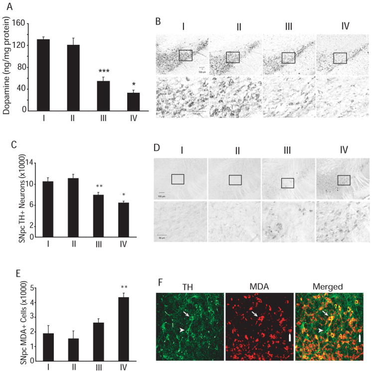
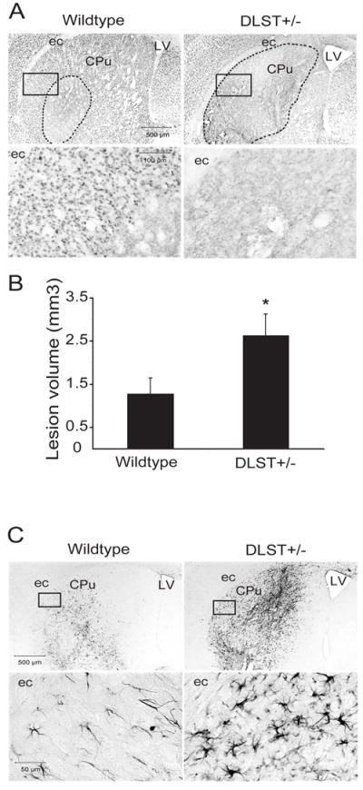
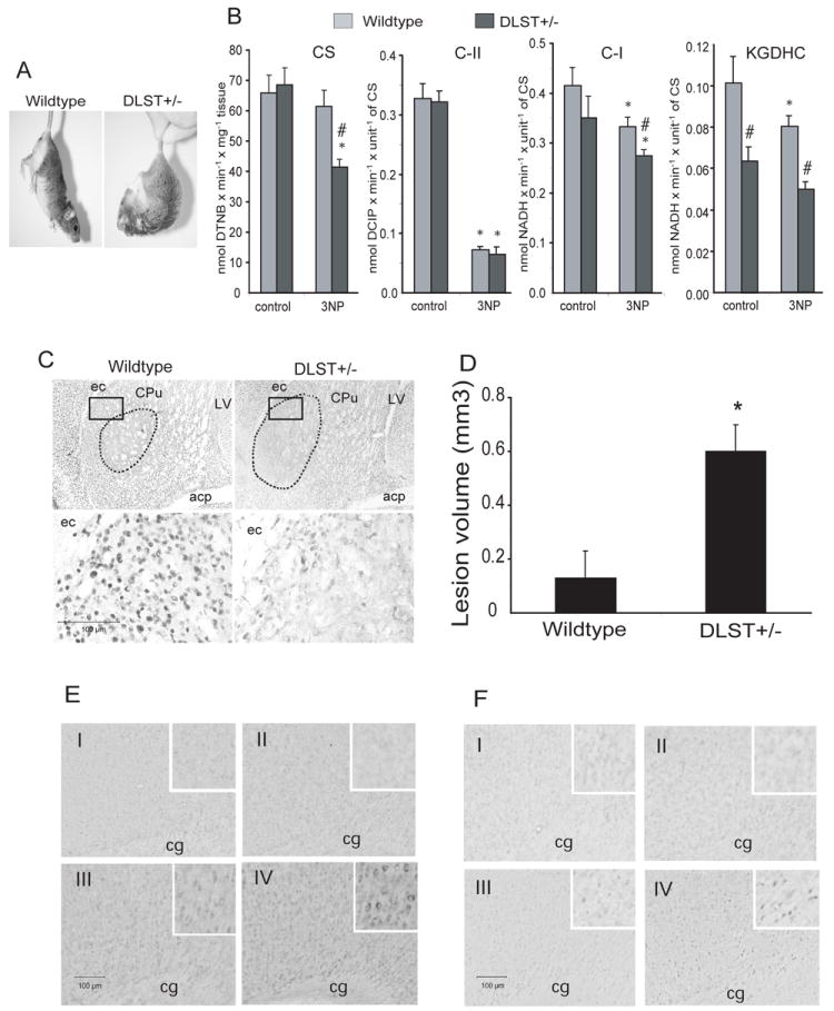
The activity of a key mitochondrial tricarboxylic acid cycle enzyme, alpha-ketoglutarate dehydrogenase complex (KGDHC), declines in many neurodegenerative diseases. KGDHC consists of three subunits. The dihydrolipoyl succinyl transferase (DLST) component is unique to KGDHC. DLST(+/-) mice showed reduced mRNA and protein levels and decreased brain mitochondrial KGDHC activity. Neurotoxic effects of mitochondrial toxins were exacerbated in DLST(+/-) mice. MPTP produced a significantly greater reduction of striatal dopamine and tyrosine hydroxylase-positive neurons in the substantia nigra pars compacta of DLST(+/-) mice. DLST deficiency enhanced the severity of lipid peroxidation in the substantia nigra after MPTP treatment. Striatal lesions induced by either malonate or 3-nitropropionic acid (3-NP) were significantly larger in DLST(+/-) mice than in wildtype controls. DLST deficiency enhanced the 3-NP inhibition of mitochondria enzymes, and 3-NP induced protein and DNA oxidations. These observations support the hypothesis that reductions in KGDHC may impair the adaptability of the brain and contribute to the pathogenesis of neurodegenerative diseases.


Figures

Figure 1
Figure 1
Gene expression and protein levels of KGDHC subunits in DLST+/- mice. A. Brain homogenates were subjected to quantitative real-time PCR to assess changes in the mRNA level of ogdh , dlst and dld subunits of KGDHC. mRNA for β-2-microglobulin (β2m) was measured in the same sample as an internal control. Values represent means ± SEM of fold changes over DLST+/+ mice from at least two independent experiments done in triplicate after normalization to β2m. * P < 0.05. B. Total protein was isolated from brains of mice and subjected to SDS-PAGE followed by Western blotting probed with antibodies against OGDH, DLST or DLD. β-actin immunoreactivity was used as an internal control. Representative blots are shown for three subunits and β-actin. C. Quantitation of protein levels of OGDH, DLST and DLD. Values represent means ± SEM of relative densities of the subunit from three independent experiments after normalization to β-actin. * P < 0.05.
Figure 2
Figure 2
KGDHC activity was reduced in brains of DLST+/- mice. A. Enzymatic activity was measured in whole brain homogenates (6 week old mice). DLST+/- brains showed significantly lower activity. B. Nitroblue tetrazolium (NBT) staining density, measured in brain sections by in situ activity staining, was lower in each region of DLST+/- brains. Abbreviations; Cb, cerebellum; Ctx, neocortex; HB, hindbrain; Hc, hippocampus; HT, hypothalamus; MB, midbrain; OB, olfactory bulb; St, striatum; Th, thalamus. Data for each group were done in at least three brains, in triplicate. * P < 0.05. All values represent means ± SEM.
Figure 3
Figure 3
KGDHC activity during development. Specific enzymatic activity was measured in triplicate. Values represent means ± SEM. * P < 0.05. n = 3-9. No significant increase in the activity after P30.
Figure 4
Figure 4
Enzyme activities in mitochondria of DLST deficient mice. A. The activity of mitochondria KGDHC isolated from DLST+/- brains was half of that from DLST+/+ brains. Other evaluated mitochondrial enzymes activities were similar in DLST+/- and DLST+/+ mitochondria. Values are means ± SEM. * p < 0.05. n = 12. B. Immunoblotting of MnSOD, GR, and GPx1 proteins in isolated mitochondria shows no difference between DLST+/- and DLST+/+. C-I, respiratory chain Complex I; COX, cytochrome oxidase; MDH, malate dehydrogenase; PDHC, pyruvate dehydrogenase complex; KGDHC, α-ketoglutarate dehydrogenase complex; ME, malic enzyme; GR, glutathione reductase; GPx1, mitochondrial glutathione peroxidase 1; MnSOD, manganese superoxide dismutase.
Figure 5
Figure 5
DLST deficiency enhances MPTP toxicity. A. DLST deficiency enhances MPTP-induced dopamine depletion in the striatum. *** p < 0.001, versus wildtype/PBS; * p < 0.05, versus wildtype/MPTP. B. Tyrosine hydroxylase (TH) immunoreactivity in the substantia nigra pars compacta (SNpc). Boxed areas on top are magnified in lower panel of photomicrographs. C. Stereological analysis of TH-immunoreactive (TH+) neurons in SNpc showing exacerbation of MPTP-induced TH+ neuron loss in the SNpc of DLST+/- mice. Values are means ± SEM. * p < 0.05, versus wildtype/MPTP; ** p < 0.01 versus wildtype/PBS. D. Malondialdehyde (MDA) immunoreactivity in the SNpc. Boxed areas on top are magnified in lower panel of photomicrographs. E. Stereological analysis of MDA-immunoreactive neurons in SNpc showing increased number of intensely stained neurons in MPTP-treated DLST-deficient mice compared to MPTP-treated wildtype control. Values are means ± SEM. ** p < 0.01 versus wildtype/MPTP. n = 10-12. I, Wildtype/PBS; II, DLST+/-/PBS; III, Wildtype/MPTP; IV, DLST+/-/MPTP. F. Confocal images of combined immunofluorescence of TH (green) and MDA (red) antisera showing co-existence (yellow) in neurons in the SNpc of a DLST+/- mouse treated with MPTP. Arrows show a TH-positive neuron with increased MDA immunoreactivity. Point bars show MDA immunostaining in a TH-negative cell. Arrowheads show a TH-immunoreactive neuron without MDA-immunoreactivity.
Figure 6
Figure 6
DLST-deficiency enhances malonate toxicity. A. NeuN-stained sections through the caudate putamen lesioned with malonate show an exacerbation of the lesion in DLST+/- mice. Broken lines show demarcation of the areas of NeuN-stained neuron loss. Boxed areas in the photomicrographs are magnified in the lower panel showing the severity of loss of NeuN-labeled neurons. B. Lesion volume analysis using stereological cavalieri method shows an increase of lesion volumes in DLST+/- mice. Values are means ± SEM. * p < 0.05. n = 10-11. C. CD40L immunoreactivity in the caudate putamen lesioned with malonate. There is marked proliferation and hypertrophy of CD40L-labeled astrocytes in DLST+/- mice. Boxed areas in the photomicrographs are magnified in the lower panel. ec= external capsule, LV= lateral ventricle, CPu= caudate putamen.
Figure 7
Figure 7
DLST-deficiency enhances 3-NP toxicity. A. Motor function in 3NP lesioned mice when suspended by the tail. After 5 days of 3-NP intoxication, the DLST+/- mouse exhibited sustained flexure of both forelimbs and hindlimbs, while the wildtype mouse showed less severe symptoms. B. Effect of 3-NP treatment on the activity of mitochondrial enzymes in striata of DLST+/- mice and their wild type littermates (see results for interpretation). * p < 0.05 compared with its respective control; # p < 0.05 compared to wildtype/3-NP. n = 3. C. NeuN-stained sections through the caudate putamen lesioned with 3-NP show an exacerbation of lesion in DLST+/- mice. Broken lines show demarcation of the areas of NeuN-stained neuron loss. Boxed areas in the photomicrographs are magnified in the lower panel to show the extent of loss of NeuN-immunoreactive neurons. ec=external capsule, LV=lateral ventricle, CPu=caudate putamen; acp=anterior commissure, posterior part. D. Lesion volumes obtained by stereological cavalieri method show an increase of lesion volumes in DLST+/- mice. Values are means ± SEM, * p < 0.05. n = 8. E. Representative photomicrographs of nitrotyrosine immunoreactivity in the cortex. Insets show the staining in neurons. F. Representative photomicrographs of 8-hydroxy-2-deoxyguanosine immunoreactivity in the cortex. Insets show the staining in neuronal nuclei. n=5 in each group. cg=cingulum. I, Wildtype/PBS; II, DLST+/-/PBS; III, Wildtype/3-NP; IV, DLST+/-/3-NP. C-I, respiratory chain Complex I; C-II, Complex II; CS, citrate synthase; KGDHC, α-ketoglutarate dehydrogenase complex.

References

    1. Alston TA, Mela L, Bright HJ. 3-Nitropropionate, the toxic substance of Indigofera, is a suicide inactivator of succinate dehydrogenase. Proc Natl Acad Sci U S A. 1977;74:3767–3771. - PMC - PubMed
    1. Beal MF, Brouillet E, Jenkins B, Henshaw R, Rosen B, Hyman BT. Age-dependent striatal excitotoxic lesions produced by the endogenous mitochondrial inhibitor malonate. J Neurochem. 1993a;61:1147–1150. - PubMed
    1. Beal MF, Ferrante RJ, Henshaw R, Matthews RT, Chan PH, Kowall NW, Epstein CJ, Schulz JB. 3-Nitropropionic acid neurotoxicity is attenuated in copper/zinc superoxide dismutase transgenic mice. J Neurochem. 1995;65:919–922. - PubMed
    1. Beal MF, Brouillet E, Jenkins BG, Ferrante RJ, Kowall NW, Miller JM, Storey E, Srivastava R, Rosen BR, Hyman BT. Neurochemical and histologic characterization of striatal excitotoxic lesions produced by the mitochondrial toxin 3-nitropropionic acid. J Neurosci. 1993b;13:4181–4192. - PMC - PubMed
    1. Brouillet E, Hantraye P, Ferrante RJ, Dolan R, Leroy-Willig A, Kowall NW, Beal MF. Chronic mitochondrial energy impairment produces selective striatal degeneration and abnormal choreiform movements in primates. Proc Natl Acad Sci U S A. 1995;92:7105–7109. - PMC - PubMed

Publication types

MeSH terms