Review
. 2009:175:393-418.
doi: 10.1016/S0079-6123(09)17526-X.

Recovery of control of posture and locomotion after a spinal cord injury: solutions staring us in the face

Affiliations
Review

Recovery of control of posture and locomotion after a spinal cord injury: solutions staring us in the face

Andy J Fong et al. Prog Brain Res. 2009.

Abstract

Over the past 20 years, tremendous advances have been made in the field of spinal cord injury research. Yet, consumed with individual pieces of the puzzle, we have failed as a community to grasp the magnitude of the sum of our findings. Our current knowledge should allow us to improve the lives of patients suffering from spinal cord injury. Advances in multiple areas have provided tools for pursuing effective combination of strategies for recovering stepping and standing after a severe spinal cord injury. Muscle physiology research has provided insight into how to maintain functional muscle properties after a spinal cord injury. Understanding the role of the spinal networks in processing sensory information that is important for the generation of motor functions has focused research on developing treatments that sharpen the sensitivity of the locomotor circuitry and that carefully manage the presentation of proprioceptive and cutaneous stimuli to favor recovery. Pharmacological facilitation or inhibition of neurotransmitter systems, spinal cord stimulation, and rehabilitative motor training, which all function by modulating the physiological state of the spinal circuitry, have emerged as promising approaches. Early technological developments, such as robotic training systems and high-density electrode arrays for stimulating the spinal cord, can significantly enhance the precision and minimize the invasiveness of treatment after an injury. Strategies that seek out the complementary effects of combination treatments and that efficiently integrate relevant technical advances in bioengineering represent an untapped potential and are likely to have an immediate impact. Herein, we review key findings in each of these areas of research and present a unified vision for moving forward. Much work remains, but we already have the capability, and more importantly, the responsibility, to help spinal cord injury patients now.


Figures

Fig. 1
Fig. 1
Brief periods of daily, high-load isometric contraction reduce the loss of muscle function after spinal cord injury. Spinal cord isolated rats (complete spinal cord transections at a mid-thoracic and a high-sacral level, plus dorsal rhizotomy performed between the two transection sites) that were administered one (SI-Stim1) or two (SI-Stim2) bouts of muscle stimulation daily exhibited less atrophy (A, muscle mass normalized to body mass) and a smaller loss of force generation capability (B, maximum tetanic tension) in the stimulated medial gastrocnemius (MG) muscle (gray bars) compared to the non-stimulated contralateral muscle (white bars, SI-C1 and SI-C2) after 30 days of treatment. Stimulation was applied at twice the minimum amplitude necessary to generate a maximum tetanic contraction. The stimulation algorithm applied a 1-s-duration, 100-Hz pulse train that was repeated every 30 s for 5 min. For Stim1, this algorithm was repeated six times over a 1-h period, with 5-min rest periods between repetitions. Stim2 also was stimulated six times, but a 9-h rest period was imposed between the third and fourth cycles. While the daily amount of stimulation was the same for both groups, rats that were stimulated twice/day versus once/day maintained muscle properties that were more similar to those of uninjured controls (black bars). Values are reported as mean±SEM. *, †, and ‡, significantly different from uninjured control (Con), from non-stimulated contralateral muscle, and from Stim1, respectively. Adapted with permission from Kim et al. (2007).
Fig. 2
Fig. 2
Monosynaptic muscle responses evoked by spinal cord stimulation are task and phase dependent. Phase-dependent modulation of the multi-segmental monosynaptic response (MMR) amplitude was observed throughout the gait cycle in the leg muscles studied in uninjured subjects. The MMR modulation pattern also was motor-task specific, differing during walking compared to running. Transcutaneous spinal cord stimulation was applied using a AgCl cathode placed on the skin overlying the T11 and T12 spinous processes during walking (left panels: 3.5 km/h) and running (right panels: 8.0 km/h). The resultant MMR responses were recorded bilaterally from selected leg muscles in eight individuals (open and shaded circles depict the left leg and right leg, respectively). Ten step cycles were analyzed for each subject. The data were discretized into 16 time bins corresponding to different periods of the step cycle, beginning with heel strike. Each data point represents the mean±SD of the MMR amplitude, reported in each individual as a percentage of the MMR amplitude recorded during standing (dashed horizontal line). All evoked potentials are recorded in millivolts. Muscles recorded: RF, rectus femoris; BF, biceps femoris; MG, medial gastrocnemius; Sol, soleus. Adapted with permission from Courtine et al. (2007).
Fig. 3
Fig. 3
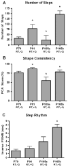
Pharmacological treatment complements robotic training in enhancing spinal locomotion. In robotically trained spinal mice (n = 8), coadministration of quipazine increased the number of steps performed (A) and improved step shape consistency (B), but did not affect step rhythm (C). After an initial period of robotic training, which ended at 79 days post-lesion (P79), increases in the number of steps performed and in step shape consistency were observed when quipazine was used to supplement training (P91). This effect was reversed when quipazine was withdrawn, demonstrating that the improvement in locomotion was attributable to the quipazine treatment (P105a). An additional bolus dose of quipazine immediately restored the pharmacologically mediated enhancement (P105b). These results suggest that quipazine and robotic training have complementary effects. Step rhythm, on the other hand, improved steadily throughout the course of robotic training, which is consistent with previous results that suggest that robotic training has a greater effect on step rhythm than quipazine. *, Significantly different from P79 (RT, −Q). RT, robotically trained; +Q, treated with quipazine; −Q, not treated with quipazine. Adapted with permission from Fong et al. (2005).
Fig. 4
Fig. 4
Diagram of rodent robotic step training and evaluation system. The rodent robotic system consists of the following major components: (A) four optical encoders, (B) four DC motors, (C) a weight-support device, (D) two 5-bar parallelogram linkages, and (E) a motor-driven treadmill. When used in an active mode, the system applies step-training algorithms that are commanded by an external motion controller. In a passive mode, the optical encoders record the trajectories of the legs during free stepping. Robotics thus enables quantitative monitoring of both training and recovery. Adapted with permission from Cai et al. (2005).
Fig. 5
Fig. 5
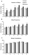
Variability in robotic step training promotes robust locomotor recovery. After 4 weeks of robotically assisted step training, complete spinal mice that were trained using a “window” control algorithm (black bar) were able to execute more steps (A), and displayed better step rhythm (B), than mice hat were trained with either a “band” algorithm (gray bar) or using traditional, continuous-assistance, “fixed” trajectory training (white bar). Contrasted with fixed trajectory training, “window” and “band” training i.e., assist-as-needed paradigms allow the hindlimb to deviate to some degree away from the nominal trained trajectory: the robotics only exert corrective action when the position of the hindlimb moves beyond a set limit, at which point a restoring force is generated (force magnitude encoded by an error-dependent velocity field). “Window” training enforces alternating interlimb coordination, whereas “band” training does not. The data suggest that the additional sensory information provided to the spinal circuitry during “window” training enhances locomotor recovery, but that interlimb coordination should be controlled when training an alternating gait. Values are reported as mean±SEM. + and #, significantly different from “fixed” and “band” training group, respectively. Adapted with permission from Cai et al. (2006).
Fig. 6
Fig. 6
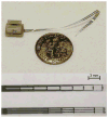
Photographs of spinal cord electrode arrays. (Top) Photograph of a spinal cord electrode array and 36-pin head connector juxtaposed against a small coin for size comparison. (Bottom) Close-up photograph of the 18-electrode contacts of a 3 × 6 electrode array with physiologically determined rostrocaudal interelectrode spacing.
Fig. 7
Fig. 7
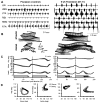
Bipedal stepping approximating that observed pre-lesion can be recovered after a complete spinal cord transection with the aid of epidural stimulation and pharmacological facilitation. EMG (A) and kinematic (B–D) data are shown for a rat before and 6 weeks after receiving a complete mid-thoracic (~T9) spinal cord transection while stepping on a treadmill at 21 cm/s. After the transection, the rat was administered quipazine and epidural stimulation. Representative stick diagram decompositions of the left hindlimb movements during the stance and swing phases of gait are shown in B. Mean waveforms of the hip, knee, and ankle joint angle for the left hindlimb are plotted for a normalized gait cycle duration in C. Each trace is an average of 15 (control) and 18 (spinal transected-trained, ST–Tr) successive steps. Horizontal bars at bottom indicate mean value of stance phase (blank) and foot drag duration (shaded). Angle–angle plots showing coupling between hip and knee (left) and knee and ankle (right) from the same data shown in C are shown in D. Filled and empty circles represent stance and swing phases of gait, respectively. Arrows indicate direction along which time is evolving. Shaded portion of the lines in C shows SEM. Adapted with permission from Gerasimenko et al. (2007).
Fig. 8
Fig. 8
A multimodal approach to spinal cord rehabilitation. It is becoming increasingly clear that combining multiple treatment paradigms can produce enhanced recovery. This diagram depicts a promising four-step approach to recovering locomotor function: (1) application of muscle stimulation to maintain normal properties of the muscles; (2) use of pharmacological treatments and epidural stimulation to recreate an electrochemical environment conducive to spinal learning; (3) administration of activity-dependent motor training to provide the appropriate cues necessary to teach the spinal cord to walk; and (4) delivery of focal epidural stimulation to refine and facilitate functional stepping patterns. All of these treatments modulate sensory input to the lumbosacral spinal circuitry, which processes the information and uses it to recover functional posture and locomotion.

Similar articles

Cited by

References

    1. Ahn SN, Guu JJ, Tobin AJ, Edgerton VR, Tillakaratne NJ. Use of c-fos to identify activity-dependent spinal neurons after stepping in intact adult rats. Spinal Cord. 2006;44:547–559. - PMC - PubMed
    1. Andersen C. Complications in spinal cord stimulation for treatment of angina pectoris. Differences in unipolar and multipolar percutaneous inserted electrodes. Acta Cardiologica. 1997;52:325–333. - PubMed
    1. Andersen JL, Mohr T, Biering-Sorensen F, Galbo H, Kjaer M. Myosin heavy chain isoform transformation in single fibres from m. vastus lateralis in spinal cord injured individuals: effects of long-term functional electrical stimulation (FES) Pflugers Archive. 1996;431:513–518. - PubMed
    1. Antri M, Barthe JY, Mouffle C, Orsal D. Long-lasting recovery of locomotor function in chronic spinal rat following chronic combined pharmacological stimulation of serotonergic receptors with 8-OHDPAT and quipazine. Neuroscience Letters. 2005;384:162–167. - PubMed
    1. Antri M, Orsal D, Barthe JY. Locomotor recovery in the chronic spinal rat: effects of long-term treatment with a 5-HT2 agonist. The European Journal of Neurosciences. 2002;16:467–476. - PubMed

Publication types

MeSH terms