A novel mouse model for the aggressive variant of NK cell and T cell large granular lymphocyte leukemia
- PMID: 19660811
- PMCID: PMC2814907
- DOI: 10.1016/j.leukres.2009.06.031
A novel mouse model for the aggressive variant of NK cell and T cell large granular lymphocyte leukemia
Abstract
Murine models of disease are vital to the understanding of pathogenesis and the development of novel therapeutics. We have previously established interleukin (IL)-15 transgenic (tg) mice that demonstrate rapid proliferation of natural killer (NK) and T cells, followed by spontaneous transformation to lethal leukemia. Herein, we have characterized this model, which has many features in common with the aggressive variants of NK and T large granular lymphocyte leukemia (LGLL) in humans. The LGLL blasts are cytolytic and produce IFN-gammaex vivo. Cytogenetic analysis revealed trisomy of chromosome 17 and/or 15. This model should provide opportunities to develop effective standard therapies for this fatal disease.
Copyright 2009 Elsevier Ltd. All rights reserved.
Conflict of interest statement
The authors do not have any conflict of interest to declare.
Figures


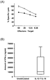
 and T LGL leukemia
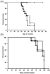
  ). (B) Kaplan Meier survival curve of SCID mice after transfer of leukemia cells to secondary recipients. Groups of 4 mice were injected intravenously with 1 × 10⁵ peripheral blood cells (-□-) or injected with same number of spleen cells (-◊-) or bone marrow cells (-Δ-) on days 0. Mice transplanted with different sources of LGL leukemic blasts showed mortality within the same period of time irrespective of the source of LGL leukemic blasts. Results shown are representative of two independent experiments that were performed.
Similar articles
-
A lineage-specific STAT5BN642H mouse model to study NK-cell leukemia.Blood. 2024 Jun 13;143(24):2474-2489. doi: 10.1182/blood.2023022655. Blood. 2024. PMID: 38498036 Free PMC article.
-
Mixed-phenotype large granular lymphocytic leukemia: a rare subtype in the large granular lymphocytic leukemia spectrum.Hum Pathol. 2018 Nov;81:96-104. doi: 10.1016/j.humpath.2018.06.023. Epub 2018 Jun 24. Hum Pathol. 2018. PMID: 29949739
-
Transformed aggressive γδ-variant T-cell large granular lymphocytic leukemia with acquired copy neutral loss of heterozygosity at 17q11.2q25.3 and additional aberrations.Eur J Haematol. 2014 Sep;93(3):260-4. doi: 10.1111/ejh.12313. Epub 2014 Apr 7. Eur J Haematol. 2014. PMID: 24635703
-
Clinical Features, Pathogenesis, and Treatment of Large Granular Lymphocyte Leukemias.Intern Med. 2017;56(14):1759-1769. doi: 10.2169/internalmedicine.56.8881. Epub 2017 Jul 15. Intern Med. 2017. PMID: 28717070 Free PMC article. Review.
-
T cell large granular lymphocyte leukemia and chronic NK lymphocytosis.Best Pract Res Clin Haematol. 2019 Sep;32(3):207-216. doi: 10.1016/j.beha.2019.06.006. Epub 2019 Jun 22. Best Pract Res Clin Haematol. 2019. PMID: 31585621 Review.
Cited by
-
Cytokines in the Pathogenesis of Large Granular Lymphocytic Leukemia.Front Oncol. 2022 Mar 10;12:849917. doi: 10.3389/fonc.2022.849917. eCollection 2022. Front Oncol. 2022. PMID: 35359386 Free PMC article. Review.
-
Cbl-b Is Upregulated and Plays a Negative Role in Activated Human NK Cells.J Immunol. 2021 Feb 15;206(4):677-685. doi: 10.4049/jimmunol.2000177. Epub 2021 Jan 8. J Immunol. 2021. PMID: 33419766 Free PMC article.
-
IL-15 Prevents the Development of T-ALL from Aberrant Thymocytes with Impaired DNA Repair Functions and Increased NOTCH1 Activation.Cancers (Basel). 2023 Jan 21;15(3):671. doi: 10.3390/cancers15030671. Cancers (Basel). 2023. PMID: 36765626 Free PMC article.
-
Out-sourcing for Trans-presentation: Assessing T Cell Intrinsic and Extrinsic IL-15 Expression with Il15 Gene Reporter Mice.Immune Netw. 2018 Feb 22;18(1):e13. doi: 10.4110/in.2018.18.e13. eCollection 2018 Feb. Immune Netw. 2018. PMID: 29503743 Free PMC article. Review.
-
A lineage-specific STAT5BN642H mouse model to study NK-cell leukemia.Blood. 2024 Jun 13;143(24):2474-2489. doi: 10.1182/blood.2023022655. Blood. 2024. PMID: 38498036 Free PMC article.
References
-
- Nakamura F, et al. Acute lymphoblastic leukemia/lymphoblastic lymphoma of natural killer (NK) lineage: quest for another NK-lineage neoplasm. Blood. 1997;89:4665–4666. - PubMed
-
- Alekshun TJ, Tao J, Sokol L. Aggressive T-cell large granular lymphocyte leukemia: a case report and review of the literature. Am J Hematol. 2007;82:481–485. - PubMed
-
- Matano S, Terahata S, Nakamura S, Kobayashi K, Sugimoto T. CD56-positive acute lymphoblastic leukemia. Acta Haematol. 2005;114:160–163. - PubMed
-
- Gentile T, et al. CD3+, CD56+ aggressive variant of large granular lymphocyte leukemia. Blood. 1994;84:2315–2321. - PubMed
-
- Johansson B, Mertens F, Mitelman F. Clinical and biological importance of cytogenetic abnormalities in childhood and adult acute lymphoblastic leukemia. Ann Med. 2004;36:492–503. - PubMed
MeSH terms
Substances
Grants and funding
LinkOut - more resources
Full Text Sources
Other Literature Sources
Miscellaneous

