Comparative Study
. 2010 Jun 1;77(2):357-66.
doi: 10.1016/j.ijrobp.2009.04.028. Epub 2009 Aug 5.

Intensity-modulated proton therapy reduces the dose to normal tissue compared with intensity-modulated radiation therapy or passive scattering proton therapy and enables individualized radical radiotherapy for extensive stage IIIB non-small-cell lung cancer: a virtual clinical study

Affiliations
Comparative Study

Intensity-modulated proton therapy reduces the dose to normal tissue compared with intensity-modulated radiation therapy or passive scattering proton therapy and enables individualized radical radiotherapy for extensive stage IIIB non-small-cell lung cancer: a virtual clinical study

Xiaodong Zhang et al. Int J Radiat Oncol Biol Phys. .

Abstract

Purpose: To compare dose volume histograms of intensity-modulated proton therapy (IMPT) with those of intensity-modulated radiation therapy (IMRT) and passive scattering proton therapy (PSPT) for the treatment of stage IIIB non-small-cell lung cancer (NSCLC) and to explore the possibility of individualized radical radiotherapy.

Methods and materials: Dose volume histograms designed to deliver IMRT at 60 to 63 Gy, PSPT at 74 Gy, and IMPT at the same doses were compared and the use of individualized radical radiotherapy was assessed in patients with extensive stage IIIB NSCLC (n = 10 patients for each approach). These patients were selected based on their extensive disease and were considered to have no or borderline tolerance to IMRT at 60 to 63 Gy, based on the dose to normal tissue volume constraints (lung volume receiving 20 Gy [V20] of <35%, total mean lung dose <20 Gy; spinal cord dose, <45 Gy). The possibility of increasing the total tumor dose with IMPT for each patient without exceeding the dose volume constraints (maximum tolerated dose [MTD]) was also investigated.

Results: Compared with IMRT, IMPT spared more lung, heart, spinal cord, and esophagus, even with dose escalation from 63 Gy to 83.5 Gy, with a mean MTD of 74 Gy. Compared with PSPT, IMPT allowed further dose escalation from 74 Gy to a mean MTD of 84.4 Gy (range, 79.4-88.4 Gy) while all parameters of normal tissue sparing were kept at lower or similar levels. In addition, IMPT prevented lower-dose target coverage in patients with complicated tumor anatomies.

Conclusions: IMPT reduces the dose to normal tissue and allows individualized radical radiotherapy for extensive stage IIIB NSCLC.


Conflict of interest statement

Conflict of Interest Notification: The authors do not have any actual or potential conflicts of interest related to this article.

Figures

Fig. 1
Fig. 1
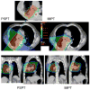
A typical case comparison between IMRT and IMPT. (a) Dose distributions for the IMRT (left) and IMPT (right) plans. Each line delineates the PTV. (b) DVHs for the IMRT plan (squares) and IMPT plan (triangles). Ips., ipsilateral; Con., contralateral.
Fig. 1
Fig. 1
A typical case comparison between IMRT and IMPT. (a) Dose distributions for the IMRT (left) and IMPT (right) plans. Each line delineates the PTV. (b) DVHs for the IMRT plan (squares) and IMPT plan (triangles). Ips., ipsilateral; Con., contralateral.
Fig. 2
Fig. 2
Comparison between IMRT and IMPT_MTD. (a) Dose distributions for the IMRT plan at 63 Gy (left) and IMPT_MTD plan at the MTD of 80 Gy (right). Each line delineates the PTV. Of note is that the esophagus was overlapped by the CTV and PTV for this patient, whereas the IMPT_MTD plan was able to reduce the esophageal dose to less than 80 Gy. (b) DVHs for the IMRT plan (squares) and IMPT_MTD plan (triangles). Ips., ipsilateral; Con., contralateral.
Fig. 2
Fig. 2
Comparison between IMRT and IMPT_MTD. (a) Dose distributions for the IMRT plan at 63 Gy (left) and IMPT_MTD plan at the MTD of 80 Gy (right). Each line delineates the PTV. Of note is that the esophagus was overlapped by the CTV and PTV for this patient, whereas the IMPT_MTD plan was able to reduce the esophageal dose to less than 80 Gy. (b) DVHs for the IMRT plan (squares) and IMPT_MTD plan (triangles). Ips., ipsilateral; Con., contralateral.
Fig. 3
Fig. 3
Comparison of IMPT with PSPT in treating an NSCLC lesion close to the spinal cord. (a) Beam arrangements for a typical PSPT plan. (b) Beam-eye view of the aperture block, CTV (green contour), PTV (blue contour), and spinal cord (red contour) for the right lateral beam. The aperture had to be edited for this beam to avoid the cord. (c) The hot region resulting from the overlap of the two beams for the PSPT plan. CTV: yellow contour. (d) Avoidance of the hot region by intensity modulation for the IMPT plan with the use of the same two beams.
Fig. 4
Fig. 4
Comparison between PSPT and IMPT. (a) Dose distributions for the PSPT (left) and IMPT (right) plans. The green lines delineate the PTV. A blown-up image of the target region for the PSPT is shown to indicate the inadequate dose coverage caused by aperture editing. (b) DVHs for the PSPT plan (squares) and IMPT plan (triangles). Ips., ipsilateral; Con., contralateral.
Fig. 4
Fig. 4
Comparison between PSPT and IMPT. (a) Dose distributions for the PSPT (left) and IMPT (right) plans. The green lines delineate the PTV. A blown-up image of the target region for the PSPT is shown to indicate the inadequate dose coverage caused by aperture editing. (b) DVHs for the PSPT plan (squares) and IMPT plan (triangles). Ips., ipsilateral; Con., contralateral.
Fig. 5
Fig. 5
IMPT bean degradation repair by depositing doses from different directions. (A) A single lateral IMPT beam passing through a textured lung-like medium with an average ρ value of 0.2 gm/cc. After passing through the target, the beam enters a uniform medium with a ρ value of 0.2 gm/cc. The degradation of the distal edge of the beam corresponds to the dose profile along the horizontal axis (the blue line in panel C). If the beam had passed through a uniform low-density medium upstream, the degradation would not have occurred. (B) The addition of AP and PA IMPT beams and use of a negative margin for the lateral beam to deposit no more than 10% of its maximum dose distal to the target. (C) The optimization process compensated for the resulting loss of target coverage by adjusting the energies and intensities of the pencils of the AP and PA beams. The resulting dose profile along the horizontal axis is indicated by the solid red line in panel C. The dashed red line is the renormalized (to 100% at the isocenter) contribution of the lateral beam to the total dose represented by the solid red line.

Similar articles

Cited by

References

    1. Curran W, Scott C, Langer C, et al. Long term benefit is observed in a phase III comparison of sequential vs concurrent chemo-radiation for patients with unresectable NSCLC: RTOG 9410. Proc Am Soc Clin Oncol. 2003:621a.
    1. Kong F, Ten Haken R, Schipper M, et al. High-dose radiation improved local tumor control and overall survival in patients with inoperable/unresectable non-small-cell lung cancer: Long-term results of a radiation dose escalation study. Int J Radiat Oncol Biol Phys. 2005;63:324–333. - PubMed
    1. Murshed H, Liu H, Liao Z, et al. Dose and volume reduction for normal lung using intensity-modulated radiotherapy for advanced-stage non-small-cell lung cancer. Int J Radiat Oncol Biol Phys. 2004;58:1258–1267. - PubMed
    1. Yom S, Liao Z, Liu H, et al. Initial evaluation of treatment-related pneumonitis in advanced- stage non-small-cell lung cancer patients treated with concurrent chemotherapy and intensity-modulated radiotherapy. Int J Radiat Oncol Biol Phys. 2007;68:94–102. - PubMed
    1. Bush DA, Slater JD, Shin BB, Cheek G, Miller DW, Slater JM. Hypofractionated proton beam radiotherapy for stage I lung cancer. Chest. 2004;126(4):1198–1203. - PubMed

Publication types

MeSH terms