. 2009 Oct 16;284(42):28865-73.
doi: 10.1074/jbc.M109.043455. Epub 2009 Aug 6.

Electrochemical measurement of electron transfer kinetics by Shewanella oneidensis MR-1

Affiliations

Electrochemical measurement of electron transfer kinetics by Shewanella oneidensis MR-1

Daniel Baron et al. J Biol Chem. .

Abstract

Shewanella oneidensis strain MR-1 can respire using carbon electrodes and metal oxyhydroxides as electron acceptors, requiring mechanisms for transferring electrons from the cell interior to surfaces located beyond the cell. Although purified outer membrane cytochromes will reduce both electrodes and metals, S. oneidensis also secretes flavins, which accelerate electron transfer to metals and electrodes. We developed techniques for detecting direct electron transfer by intact cells, using turnover and single turnover voltammetry. Metabolically active cells attached to graphite electrodes produced thin (submonolayer) films that demonstrated both catalytic and reversible electron transfer in the presence and absence of flavins. In the absence of soluble flavins, electron transfer occurred in a broad potential window centered at approximately 0 V (versus standard hydrogen electrode), and was altered in single (DeltaomcA, DeltamtrC) and double deletion (DeltaomcA/DeltamtrC) mutants of outer membrane cytochromes. The addition of soluble flavins at physiological concentrations significantly accelerated electron transfer and allowed catalytic electron transfer to occur at lower applied potentials (-0.2 V). Scan rate analysis indicated that rate constants for direct electron transfer were slower than those reported for pure cytochromes (approximately 1 s(-1)). These observations indicated that anodic current in the higher (>0 V) window is due to activation of a direct transfer mechanism, whereas electron transfer at lower potentials is enabled by flavins. The electrochemical dissection of these activities in living cells into two systems with characteristic midpoint potentials and kinetic behaviors explains prior observations and demonstrates the complementary nature of S. oneidensis electron transfer strategies.


Figures
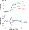
FIGURE 1.
FIGURE 1.
A, cyclic voltammetry (1 mV/s) of S. oneidensis MR-1 after 96 h of growth on planar 2-cm2 electrodes at +0. 24 V versus SHE. After the addition of cells, the initial levels of flavins were 0.2 μm, unless riboflavin was provided at inoculation as shown. B, first derivative plots of data in A, showing midpoint potentials of initial potential increase and similar slopes in higher potential regions.
FIGURE 2.
FIGURE 2.
Attached S. oneidensis MR-1 protein after 96 h of growth on planar electrodes at +0.24 V versus SHE. The ratio of attached biomass to limiting current produced in cyclic voltammetry is shown on the right axis. The error bars show standard deviations from three independent reactors.
FIGURE 3.
FIGURE 3.
A and B, scanning electron micrographs of electrodes before (A) and after (B) preparation of S. oneidensis MR-1 films. C, confocal microscopy (maximum projections, top view) of similar electrodes after exposure to cells and (D) after washing five times to remove loosely attached cells.
FIGURE 4.
FIGURE 4.
A, cyclic voltammetry (1 mV/s) of electrodes with S. oneidensis MR-1 cell films under starvation conditions (red trace), 30 min after the addition of 20 mm lactate as an electron donor (blue trace), and 30 min after the addition of 1 μm riboflavin to lactate-amended cells (black trace). B and C, identical experiments for thin films of mutants containing deletions in omcA and mtrC, respectively. Two sweeps were performed for each CV, and the second is shown. WT, wild type.
FIGURE 5.
FIGURE 5.
Base line-subtracted cyclic voltammetry data (1 mV/s) showing reversible electron transfer by adsorbed S. oneidensis MR-1 cell films, in black. The data from ΔmtrC is in red, that from ΔomcA is in blue, and that from ΔmtrComcA is in green. The current vales are normalized to represent all electrodes containing 1 μg of protein/cm2. The two similarly colored traces show data from two independent replicates, showing variability between films. Two sweeps were performed for each CV, and the second is shown. WT, wild type.
FIGURE 6.
FIGURE 6.
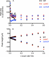
A, integrated peak area for cathodic (open symbols) and anodic (closed symbols) for wild type and mutant cell films as a function of scan rate. B, relationship between peak height and scan rate for representative S. oneidensis wild type and mutant cell films for both anodic and cathodic peaks. WT, wild type.
FIGURE 7.
FIGURE 7.
Representative data showing the peak potential separation versus log (scan rate) for S. oneidensis MR-1 cell films. The solid line shows an example of a fit for a wild type film, where the data were fit to a k0 of 2 s−1. The dotted lines show peak separation ranges consistent with 0.1, 1, and 10 s−1, respectively.

References

    1. Richardson D. J. (2000) Microbiology 146, 551–571 - PubMed
    1. Nealson K. H., Saffarini D. (1994) Annu. Rev. Microbiol. 48, 311–343 - PubMed
    1. Lovley D. R., Holmes D. E., Nevin K. P. (2004) Adv. Microb. Physiol. 49, 219–286 - PubMed
    1. Gralnick J. A., Newman D. K. (2007) Mol. Microbiol. 65, 1–11 - PMC - PubMed
    1. Nealson K. H., Scott J. (2005) in The Prokaryotes, pp. 1133–1151, Springer-Verlag New York Inc., New York

Publication types

LinkOut - more resources