. 2009 Nov;155(Pt 11):3532-3543.
doi: 10.1099/mic.0.029199-0. Epub 2009 Aug 6.

Rapid and spontaneous loss of phthiocerol dimycocerosate (PDIM) from Mycobacterium tuberculosis grown in vitro: implications for virulence studies

Affiliations

Rapid and spontaneous loss of phthiocerol dimycocerosate (PDIM) from Mycobacterium tuberculosis grown in vitro: implications for virulence studies

Pilar Domenech et al. Microbiology (Reading). 2009 Nov.

Abstract

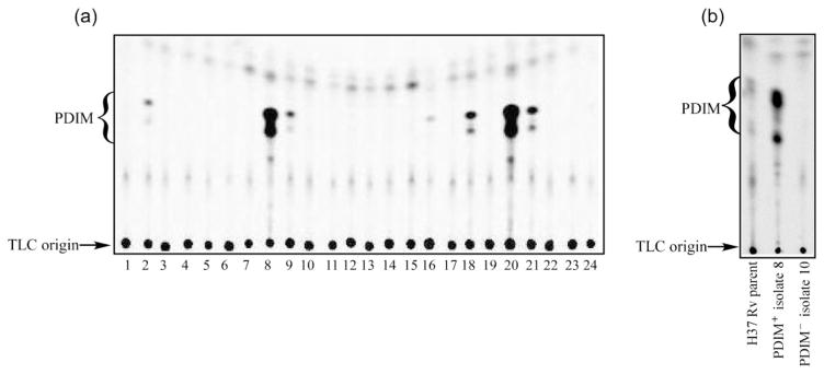
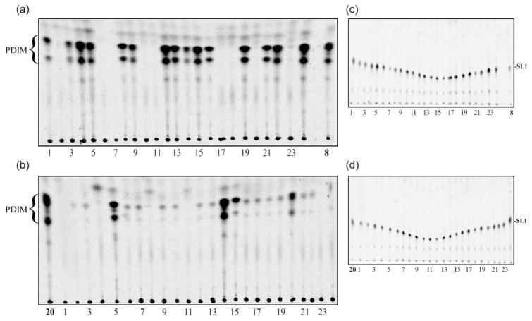
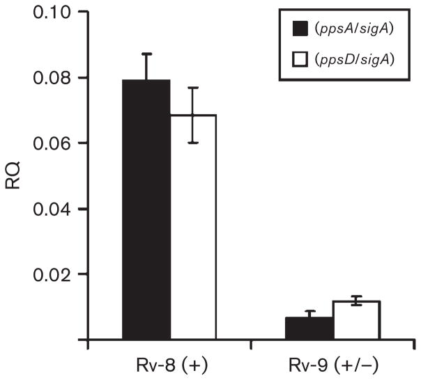
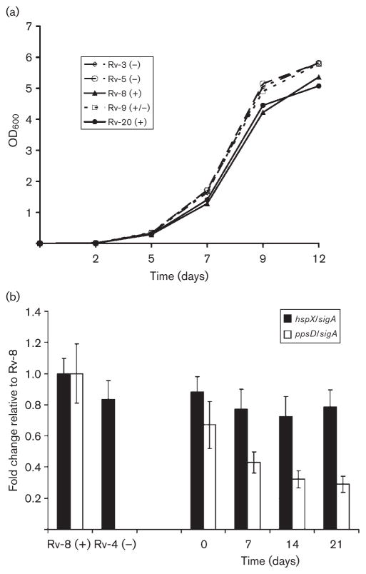
Isolated in vitro more than half a century ago, the H37Rv strain of Mycobacterium tuberculosis still remains the strain of choice for the majority of laboratories conducting in vivo studies of TB pathogenesis. In this report we reveal that H37Rv is highly prone to losing the ability to synthesize the cell wall lipid phthiocerol dimycocerosate (PDIM) during extended periods of in vitro culture. In addition, H37Rv stocks that have been held in vitro for even a short length of time should be thought of as a heterogeneous population of PDIM-positive and PDIM-negative cell types. We demonstrate that after weekly subculture of PDIM-positive isolates over a period of 20 weeks, the proportion of PDIM-negative cells rises above 30 %. That PDIM biosynthesis is negatively selected in vitro is evident from the broad range of mutation types we observe within cultures originating from a single PDIM-positive parental clone. Moreover, the appearance of these multiple mutation types coupled with an enhanced growth rate of PDIM-negative bacteria ensures that 'PDIM-less' clones rapidly dominate in vitro cultures. It has been known for almost a decade that strains of M. tuberculosis that lack PDIM are severely attenuated during in vivo infection. Therefore, the loss of PDIM raises a very serious issue in regard to the interpretation of putative virulence factors where heterogeneous parental cultures are potentially being compared in vivo to recombinant clones isolated within a PDIM-negative background. It is essential that researchers undertaking in vivo virulence studies confirm the presence of PDIM within all recombinant clones and the parental strains they are derived from.


Figures

Fig. 1
Fig. 1
PDIM-deficient mutants arise spontaneously in vitro in the absence of additional selection pressure. (a) TLC of apolar lipid extracts prepared for 24 individual clones derived from an untreated M. tuberculosis H37Rv (ATCC 27294) liquid culture. Cultures were radiolabelled with [1-14C]propionic acid prior to extraction and the TLC was developed three times in petroleum ether/ethyl acetate (98 : 2, v/v). (b) TLC of apolar lipid extracts prepared from the parental H37Rv (ATCC 27294) stock and representative PDIM-positive (no. 8) and PDIM-negative (no. 10) subclones identified in (a).
Fig. 2
Fig. 2
Rapid loss of PDIM during serial passage of H37Rv in vitro. After weekly subculture over a period of 20 weeks, apolar lipid extracts were prepared from 24 subclones derived from two of the strongly PDIM-positive cultures identified in Fig. 1(a). Liquid cultures were radiolabelled with [1-14C]propionic acid prior to extraction and the TLCs for PDIM detection were developed three times in petroleum ether/ethyl acetate (98 : 2, v/v) (panels a, b). Subclones derived from isolates 8 (panels a, c) and 20 (panels b, d) are shown and the parental isolates are indicated. The same TLC plates were also analysed for sulfolipid-1 (SL-1) (panels c, d). The solvent system in this case was chloroform/methanol/water (100 : 14 : 0.8, by vol.).
Fig. 3
Fig. 3
Repression of genes involved in phthiocerol biosynthesis is associated with a defect in PDIM production in vitro. qRT-PCR analysis was carried out on cDNA samples prepared from the PDIM-positive isolate 8 (Rv-8, PDIM+) and the weakly positive isolate 9 (Rv-9, PDIM+/−). Relative expression data (RQ, or relative quantity) were obtained using primers specific for the ppsA and ppsD genes normalized to the sigA housekeeping gene. The cDNA used in these experiments was prepared from RNA samples obtained independently of the samples analysed via microarray (Table 4) and serves to confirm the array data. Each cDNA sample was assayed twice (in triplicate) and standard deviations are indicated.
Fig. 4
Fig. 4
Loss of PDIM confers a growth advantage in vitro. (a) In vitro growth curves for two PDIM-positive isolates (8 and 20; solid lines), a weakly positive isolate (9) and two PDIM-negative isolates (3 and 5). Data obtained from one of two independent biological replicates are presented. (b, left) qRT-PCR analysis of genomic DNA prepared from PDIM-positive isolate 8 (Rv-8) and PDIM-negative isolate 4 (Rv-4). Data obtained with primers specific for the hspX and ppsD genes were normalized to sigA. The normalized values are plotted relative to the wild-type PDIM-positive strain (no. 8). These data confirm that ppsD is absent from the PDIM-negative strain. (b, right) The proportion of isolate 8 (PDIM+) and isolate 4 (PDIM) when grown in mixed culture was assayed by qRT-PCR over a period of 21 days. Primers specific for hspX were used for quantification of PDIM+ and PDIM isolates in the mixture, whilst ppsD primers were specific for the PDIM+ isolate. Data are normalized to sigA and the results are expressed relative to Rv-8. Standard deviations are indicated in (b).

References

    1. Andreu N, Gibert I. Cell population heterogeneity in Mycobacterium tuberculosis H37Rv. Tuberculosis (Edinb) 2008;88:553–559. - PubMed
    1. Applied Biosystems. Real-Time PCR Systems: Chemistry Guide. Foster City, CA: Applied Biosystems; 2005. Part Number 4348358 Rev. E edn.
    1. Astarie-Dequeker C, Le Guyader L, Malaga W, Seaphanh FK, Chalut C, Lopez A, Guilhot C. Phthiocerol dimycocerosates of M. tuberculosis participate in macrophage invasion by inducing changes in the organization of plasma membrane lipids. PLoS Pathog. 2009;5:e1000289. - PMC - PubMed
    1. Azad AK, Sirakova TD, Rogers LM, Kolattukudy PE. Targeted replacement of the mycocerosic acid synthase gene in Mycobacterium bovis BCG produces a mutant that lacks mycosides. Proc Natl Acad Sci U S A. 1996;93:4787–4792. - PMC - PubMed
    1. Azad AK, Sirakova TD, Fernandes ND, Kolattukudy PE. Gene knockout reveals a novel gene cluster for the synthesis of a class of cell wall lipids unique to pathogenic mycobacteria. J Biol Chem. 1997;272:16741–16745. - PubMed

Publication types

MeSH terms