. 2009 Oct 15;180(8):720-30.
doi: 10.1164/rccm.200904-0573OC. Epub 2009 Aug 6.

Allergic sensitization through the airway primes Th17-dependent neutrophilia and airway hyperresponsiveness

Affiliations

Allergic sensitization through the airway primes Th17-dependent neutrophilia and airway hyperresponsiveness

Rhonda H Wilson et al. Am J Respir Crit Care Med. .

Abstract

Rationale: In humans, immune responses to inhaled aeroallergens develop in the lung and draining lymph nodes. Many animal models of asthma bypass this route and instead use intraperitoneal injections of allergen using aluminum hydroxide as an adjuvant.

Objectives: We investigated whether allergic sensitization through the airway elicits immune responses qualitatively different than those arising in the peritoneum.

Methods: Mice were sensitized to allergen through the airway using low-dose LPS as an adjuvant, or through the peritoneum using aluminum hydroxide as an adjuvant. After a single allergen challenge, ELISA and flow cytometry were used to measure cytokines and leukocyte subsets. Invasive measurements of airway resistance were used to measure allergen-induced airway hyperreactivity (AHR).

Measurements and main results: Sensitization through the peritoneum primed strong Th2 responses and eosinophilia, but not AHR, after a single allergen challenge. By contrast, allergic sensitization through the airway primed only modest Th2 responses, but strong Th17 responses. Th17 cells homed to the lung and released IL-17 into the airway on subsequent encounter with inhaled allergen. As a result, these mice developed IL-17-dependent airway neutrophilia and AHR. This AHR was neutrophil-dependent because it was abrogated in CXCR2-deficient mice and also in wild-type mice receiving a neutrophil-depleting antibody. Individually, neither IL-17 nor ongoing Th2 responses were sufficient to confer AHR, but together they acted synergistically to promote neutrophil recruitment, eosinophil recruitment and AHR.

Conclusions: Allergic sensitization through the airway primes modest Th2 responses but strong Th17 responses that promote airway neutrophilia and acute AHR. These findings support a causal role for neutrophils in severe asthma.


Figures
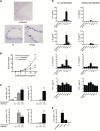
Figure 1.
Figure 1.
Method of allergen sensitization affects biologic and pathophysiologic responses to allergen challenge. C57BL/6 mice were sensitized to ovalbumin (OVA) through the peritoneum using aluminum hydroxide as an adjuvant, or through the airway using LPS as an adjuvant. All animals were challenged on a single occasion with aerosolized OVA and analyzed 48 hours post challenge. (A) Alcian blue staining of mucus-producing cells. (B) Invasive measurements of methacholine-induced airway resistance. (C) Comparison of total leukocytes and individual leukocyte subsets in the bronchoalveolar lavage (BAL) of airway-sensitized and intraperitoneally sensitized mice. (D) Cytokine levels in the BAL at various times post challenge. (E) Time course of neutrophil recruitment to the airway after sensitization only. *P ≤ 0.05; **P ≤ 0.01; ***P ≤ 0.001 denote significance between intraperitoneally sensitized and airway-sensitized groups of mice (n = 6–13 mice per group).
Figure 2.
Figure 2.
Impact of LPS on ovalbumin (OVA) sensitization through the airway and peritoneum. C57BL/6 mice were sensitized through the airway or peritoneum using the indicated levels of LPS as adjuvant. In addition to mice receiving OVA together with LPS, control groups include mice receiving OVA alone or LPS alone. For intraperitoneal injections, one group received aluminum hydroxide as a control adjuvant. All animals were challenged on a single occasion with aerosolized OVA and analyzed 48 hours post challenge (n = 7 mice per group).
Figure 3.
Figure 3.
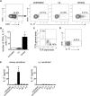
Sensitization through the airway selectively primes IL-17 cells. Mice were sensitized through the airway or peritoneum and harvested 1 week after the second sensitization. Leukocytes were prepared from lungs of these mice, as well as from untreated control animals, and analyzed by flow cytometry. (A) Representative individual flow plots and gating strategies are shown for CD4+ T cells, as well as for IL-17+ cells within the CD4+ gate. (B) Bar histograms depict total number of CD4+ IL-17+ T cells of untreated, and of airway-sensitized and intraperitoneally sensitized mice. (C) αβ and δγ TCR staining of cells within IL-17+ cell gate of airway-sensitized mice. (D) Analysis of IL-17+ T cells in the spleen of airway-sensitized mice. (E) Levels of IL-17 in the airway at various times post ovalbumin challenge. *P ≤ 0.05 denotes significance between intraperitoneally sensitized and airway-sensitized groups, each having three pools of mice with three mice in each pool. Data shown represent the results of one of two similar experiments.
Figure 4.
Figure 4.
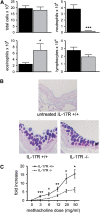
Airway neutrophilia and airway hyperreactivity is dependent on signaling responses to IL-17. Mice were sensitized twice through the airway and analyzed 48 hours after a single ovalbumin (OVA) challenge. (A) Total leukocytes and individual subsets in the bronchoalveolar lavage of wild-type (WT) and Il-17ra−/− mice. Data shown are compiled from three similar experiments (n = 24 mice per group). Solid columns represent IL-17R+/+, shaded columns represent IL-17R−/−. (B) Alcian blue/periodic acid-Schiff staining of mucus-producing cells. (C) Invasive measurements of lung resistance 48 hours post OVA challenge in WT and IL-17ra−/− mice (n = 16 mice per group). *P ≤ 0.05; **P ≤ 0.01; ***P ≤ 0.001. The data shown are compiled from two similar experiments yielding similar results.
Figure 5.
Figure 5.
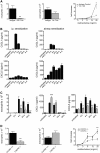
Neutrophil recruitment to the airway is required for airway hyperreactivity (AHR). (A) Mice were sensitized twice through the airway and given anti–GR-1 antibody or isotype control antibody 6 hours before ovalbumin (OVA) challenge. The accumulation of neutrophils and eosinophils in the airway is shown, as well as AHR (n = 8 mice per group). (B) C57BL/6 mice were sensitized twice through the airway or peritoneum as indicated and harvested at various times post OVA challenge. CXCL1 and CXCL5 levels in the bronchoalveolar lavage (BAL) were measured by ELISA. (C) C57BL/6 and Il-17ra−/− mice were sensitized through the airway and harvested at various times post challenge. CXCL1 and CXCL5 levels in the BAL were measured by ELISA. Solid columns represent IL-17R+/+; shaded columns represent IL-17R−/−. (D) Wild-type BALB/c and genetically matched Cxcr2−/− mice were sensitized twice through the airway. Levels of neutrophils, eosinophils, and AHR were measured at 48 hours post OVA challenge. (n = 21 mice per group) *P ≤ 0.05; **P ≤ 0.01; ***P ≤ 0.001.
Figure 6.
Figure 6.
Airway administration of chemokines induces neutrophilia, but not airway hyperreactivity. Mice were sensitized through the airway or peritoneum and challenged on a single occasion with aerosolized ovalbumin (OVA). Where indicated, intraperitoneally sensitized mice received airway delivery of either CXCL5 or CXCL1 at 4 hours post OVA challenge. All animals were harvested 48 hours later for levels of bronchoalveolar lavage neutrophils and airway hyperreactivity. ***P ≤ 0.001.
Figure 7.
Figure 7.
IL-17 and Th2 responses act synergistically to promote airway neutrophilia and airway hyperreactivity (AHR). All groups of C57BL/6 mice received intraperitoneal sensitizations to prime Th2 immunity, and some of these mice were also challenged with aerosolized ovalbumin (OVA). Where indicated, mice also received airway delivery of exogenous IL-17 or IL-17F. (A) Neutrophil and eosinophil recruitment to the airway 48 hours post OVA challenge. (B) Methacholine-induced airway resistance in mice receiving IL-17 (left panel) or IL-17F (right panel). Data shown represent the results of three similar experiments. (C) Wild-type and Stat6−/− mice were sensitized through the airway, challenged with OVA, and assessed for eosinophil and neutrophil accumulation in the airway, and for methacholine-induced airway hyperractivity at 48 hours post challenge. Data shown represent the results of two similar experiments (n = 10 mice per group). *P ≤ 0.05; **P ≤ 0.01; ***P ≤ 0.001.

References

    1. Herrick CA, Bottomly K. To respond or not to respond: T cells in allergic asthma. Nat Rev Immunol 2003;3:405–412. - PubMed
    1. Larche M, Robinson DS, Kay AB. The role of T lymphocytes in the pathogenesis of asthma. J Allergy Clin Immunol 2003;111:450–463, quiz 464. - PubMed
    1. Holgate ST. Novel targets of therapy in asthma. Curr Opin Pulm Med 2009;15:63–71. - PubMed
    1. Molet S, Hamid Q, Davoine F, Nutku E, Taha R, Page N, Olivenstein R, Elias J, Chakir J. IL-17 is increased in asthmatic airways and induces human bronchial fibroblasts to produce cytokines. J Allergy Clin Immunol 2001;108:430–438. - PubMed
    1. Barnes PJ. New molecular targets for the treatment of neutrophilic diseases. J Allergy Clin Immunol 2007;119:1055–1062, quiz 1063–1064. - PubMed

Publication types