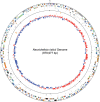
Analysis of complete genome sequence of Neorickettsia risticii: causative agent of Potomac horse fever
- PMID: 19661282
- PMCID: PMC2764437
- DOI: 10.1093/nar/gkp642
Analysis of complete genome sequence of Neorickettsia risticii: causative agent of Potomac horse fever
Abstract
Neorickettsia risticii is an obligate intracellular bacterium of the trematodes and mammals. Horses develop Potomac horse fever (PHF) when they ingest aquatic insects containing encysted N. risticii-infected trematodes. The complete genome sequence of N. risticii Illinois consists of a single circular chromosome of 879 977 bp and encodes 38 RNA species and 898 proteins. Although N. risticii has limited ability to synthesize amino acids and lacks many metabolic pathways, it is capable of making major vitamins, cofactors and nucleotides. Comparison with its closely related human pathogen N. sennetsu showed that 758 (88.2%) of protein-coding genes are conserved between N. risticii and N. sennetsu. Four-way comparison of genes among N. risticii and other Anaplasmataceae showed that most genes are either shared among Anaplasmataceae (525 orthologs that generally associated with housekeeping functions), or specific to each genome (>200 genes that are mostly hypothetical proteins). Genes potentially involved in the pathogenesis of N. risticii were identified, including those encoding putative outer membrane proteins, two-component systems and a type IV secretion system (T4SS). The bipolar localization of T4SS pilus protein VirB2 on the bacterial surface was demonstrated for the first time in obligate intracellular bacteria. These data provide insights toward genomic potential of N. risticii and intracellular parasitism, and facilitate our understanding of PHF pathogenesis.
Figures


Similar articles
-
Neorickettsia risticii surface-exposed proteins: proteomics identification, recognition by naturally-infected horses, and strain variations.Vet Res. 2011 Jun 2;42(1):71. doi: 10.1186/1297-9716-42-71. Vet Res. 2011. PMID: 21635728 Free PMC article.
-
Isolation and Molecular Analysis of a Novel Neorickettsia Species That Causes Potomac Horse Fever.mBio. 2020 Feb 25;11(1):e03429-19. doi: 10.1128/mBio.03429-19. mBio. 2020. PMID: 32098825 Free PMC article.
-
An Ecotype of Neorickettsia risticii Causing Potomac Horse Fever in Canada.Appl Environ Microbiol. 2016 Sep 16;82(19):6030-6. doi: 10.1128/AEM.01366-16. Print 2016 Oct 1. Appl Environ Microbiol. 2016. PMID: 27474720 Free PMC article.
-
Potomac Horse Fever.Vet Clin North Am Equine Pract. 2023 Apr;39(1):37-45. doi: 10.1016/j.cveq.2022.11.010. Epub 2023 Feb 1. Vet Clin North Am Equine Pract. 2023. PMID: 36737286 Review.
-
Historical aspects of Potomac horse fever in Ontario (1924-2010).Can Vet J. 2013 Jun;54(6):565-72. Can Vet J. 2013. PMID: 24155447 Free PMC article. Review.
Cited by
-
Ultrastructure and localization of Neorickettsia in adult digenean trematodes provides novel insights into helminth-endobacteria interaction.Parasit Vectors. 2017 Apr 13;10(1):177. doi: 10.1186/s13071-017-2123-7. Parasit Vectors. 2017. PMID: 28407790 Free PMC article.
-
Proteomic analysis of Neorickettsia sennetsu surface-exposed proteins and porin activity of the major surface protein P51.J Bacteriol. 2010 Nov;192(22):5898-905. doi: 10.1128/JB.00632-10. Epub 2010 Sep 10. J Bacteriol. 2010. PMID: 20833807 Free PMC article.
-
Real-Time PCR Differential Detection of Neorickettsia findlayensis and N. risticii in Cases of Potomac Horse Fever.J Clin Microbiol. 2022 Jul 20;60(7):e0025022. doi: 10.1128/jcm.00250-22. Epub 2022 Jun 13. J Clin Microbiol. 2022. PMID: 35695520 Free PMC article.
-
Neorickettsia risticii surface-exposed proteins: proteomics identification, recognition by naturally-infected horses, and strain variations.Vet Res. 2011 Jun 2;42(1):71. doi: 10.1186/1297-9716-42-71. Vet Res. 2011. PMID: 21635728 Free PMC article.
-
Detection of Neorickettsia risticii, the agent of Potomac horse fever, in horses from Rio de Janeiro, Brazil.Sci Rep. 2020 Apr 29;10(1):7208. doi: 10.1038/s41598-020-64328-2. Sci Rep. 2020. PMID: 32350359 Free PMC article.
References
-
- Knowles RC, Anderson CW, Shipley WD, Whitlock RH, Perry BD, Davidson JD. Proceedings of the 29th Annual Convention of the American Association of Enquine Practitioners. Lexington, KY: American Association of Equine Practitioners; 1984. pp. 353–357.
-
- Rikihisa Y, Perry B, Cordes D. Rickettsial link with acute equine diarrhoea. Vet. Rec. 1984;115:390. - PubMed
-
- Holland CJ, Ristic M, Cole AI, Johnson P, Baker G, Goetz T. Isolation, experimental transmission, and characterization of causative agent of Potomac horse fever. Science. 1985;227:522–524. - PubMed
Publication types
MeSH terms
Substances
Associated data
- Actions
Grants and funding
LinkOut - more resources
Full Text Sources
Other Literature Sources
Molecular Biology Databases

