. 2009;18(3):665-75.
doi: 10.3233/JAD-2009-1179.

Neuropathologic studies of the Baltimore Longitudinal Study of Aging (BLSA)

Affiliations

Neuropathologic studies of the Baltimore Longitudinal Study of Aging (BLSA)

Richard J O'Brien et al. J Alzheimers Dis. 2009.

Abstract

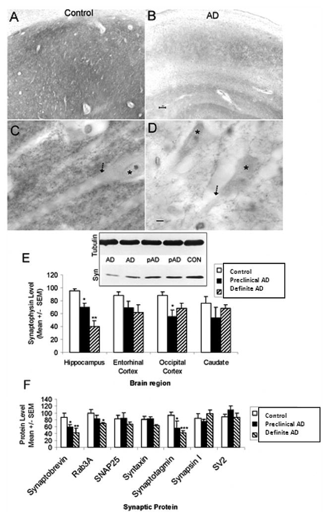
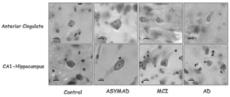
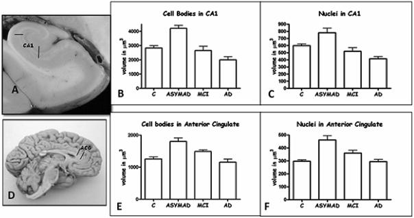
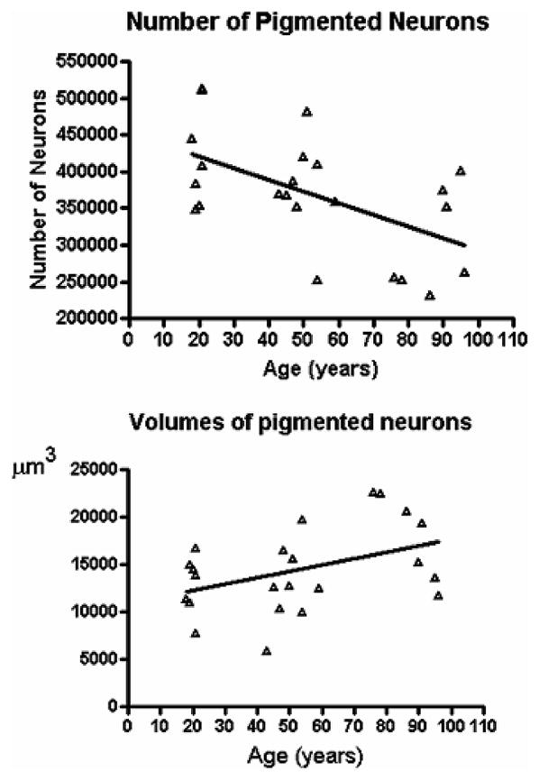
The Baltimore Longitudinal Study of Aging (BLSA) was established in 1958 and is one the oldest prospective studies of aging in the USA and the world. The BLSA is supported by the National Institute of Aging (NIA) and its mission is to learn what happens to people as they get old and how to sort out changes due to aging from those due to disease or other causes. In 1986, an autopsy program combined with comprehensive neurologic and cognitive evaluations was established in collaboration with the Johns Hopkins University Alzheimer's Disease Research Center (ADRC). Since then, 211 subjects have undergone autopsy. Here we review the key clinical neuropathological correlations from this autopsy series. The focus is on the morphological and biochemical changes that occur in normal aging, and the early neuropathological changes of neurodegenerative diseases, especially Alzheimer's disease (AD). We highlight the combined clinical, pathologic, morphometric, and biochemical evidence of asymptomatic AD, a state characterized by normal clinical evaluations in subjects with abundant AD pathology. We conclude that in some individuals, successful cognitive aging results from compensatory mechanisms that occur at the neuronal level (i.e., neuronal hypertrophy and synaptic plasticity) whereas a failure of compensation may culminate in disease.


Figures

Fig. 1
Fig. 1
Abnormalities occur in selective synaptic proteins in preclinical/asymptomatic AD and AD brain. (A-D) Immunohistochemical localization (brown labeling) of the presynaptic vesicle protein synaptophysin in human control (A,C) and AD (B,D) CA1 region of hippocampus with cresyl violet counterstaining. In controls, the neuropil of CA1 is highly enriched in synaptophysin immunoreactivity (A) which can be seen as discrete small particle-like boutons that decorate the surfaces of dendrites (arrow) of CA1 pyramidal neurons (asterisk). In the AD brain, the CA1 region is severely depleted of synaptophysin immunoreactivity (B) and the labeling of synaptophysin-positive presynaptic terminals on dendrites (arrow) of CA1 neurons (asterisk) and in the surrounding neuropil is reduced markedly. Scale bars = 10 μm. (E) Graph showing the results of immunoblot experiments of synaptophysin protein levels in different brain regions of control, preclinical/asymptomatic AD (pAD), and definite AD cases. A representative blot for synaptophysin (Syn) is shown with β-tubulin immunoreactivity serving as a protein loading control. Significant differences in synaptophysin levels from control are indicated by single asterisk (p < 0.05) or double asterisk (p < 0.0001). For more about this study see Sze et al., 1997. (F) Graph showing the result of immunoblot experiments of different presynaptic vesicle exocytosis proteins (synaptobrevin, Rab3A, synaptotagmin, synapsin I, and SV2) and presynaptic plasma membrane proteins (SNAP25 and syntaxin) in CA1 hippocampus of control, preclinical/asymptomatic AD, and definite AD cases. Significant differences in protein levels from control are indicated by single asterisk (p < 0.05), double asterisk (p < 0.01) or triple asterisk (p < 0.005). For more about this study see Sze et al., 2000 [25]. (Colours are visible in the electronic version of the article at www.iospress.nl.)
Fig. 2
Fig. 2
This panel illustrates representative neurons in the anterior cingulate (top row) and CA1-hippocampus (bottom row) in each study group. Note the neuronal hypertrophy in ASYMAD. Nissl stain sections. All magnification bars are 10 μm. (Colours are visible in the electronic version of the article at www.iospress.nl.)
Fig. 3
Fig. 3
A and D illustrate the anatomical sites from where tissue sections were taken: CA1-hippocampus (A) and anterior cingulate (D). The histograms (B, C, E, and F) show the mean volumes of neuronal cell bodies and nuclei. In CA1, comparing ASYMAD versus controls, the mean volume of neuronal cell bodies is 48.7% larger (p < 0.0001) in ASYMAD (B), and the nuclear volume 30.4% (p < 0.001) larger in ASYMAD (C). In anterior cingulate gyrus, the cell bodies are 43.5% (p < 0.0001) larger (E) and nuclei 55.5% (p < 0.0001) larger (F) in ASYMAD compared to controls. The bars correspond to the standard errors of the mean.
Fig. 4
Fig. 4
A) Linear regression of the total number of pigmented neurons in one side of the substantia nigra versus age is significant (p = 0.0060) with R2 = 0.337; CV = 0.20. B) Linear regression of the mean volume of the pigmented neurons in the substantia nigra versus age (p = 0.046) withR2 = 0.18; CV = 0.31.

References

    1. Shock NW, Greulich RC, Costa PTJ, Andres R, Lakatta EG, Arenberg D, Tobin JD. Normal Human Aging: The Baltimore Longitudinal Study of Aging. U.S. Government Printing Office; Washington, D.C.: 1984.
    1. Kawas C, Gray S, Brookmeyer R, Fozard J, Zonderman A. Age-specific incidence rates of Alzheimer’s disease: the Baltimore Longitudinal Study of Aging. Neurology. 2000;54:2072–2077. - PubMed
    1. Hughes CP, Berg L, Danziger WL, Coben LA, Martin RL. A new clinical scale for the staging of dementia. Br J Psychiatry. 1982;140:566–572. - PubMed
    1. Morris JC, Ernesto C, Schafer K, Coats M, Leon S, Sano M, Thal LJ, Woodbury P. Clinical dementia rating training and reliability in multicenter studies: the Alzheimer’s Disease Cooperative Study experience. Neurology. 1997;48:1508–1510. - PubMed
    1. Silverman JM, Breitner JC, Mohs RC, Davis KL. Reliability of the family history method in genetic studies of Alzheimer’s disease and related dementias. Am J Psychiatry. 1986;143:1279–1282. - PubMed

Publication types

MeSH terms