

Architecture and secondary structure of an entire HIV-1 RNA genome
- PMID: 19661910
- PMCID: PMC2724670
- DOI: 10.1038/nature08237
Architecture and secondary structure of an entire HIV-1 RNA genome
Abstract
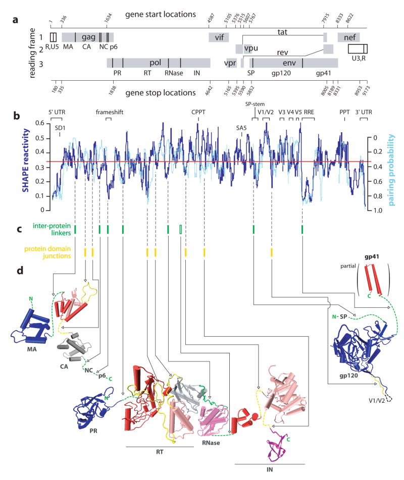
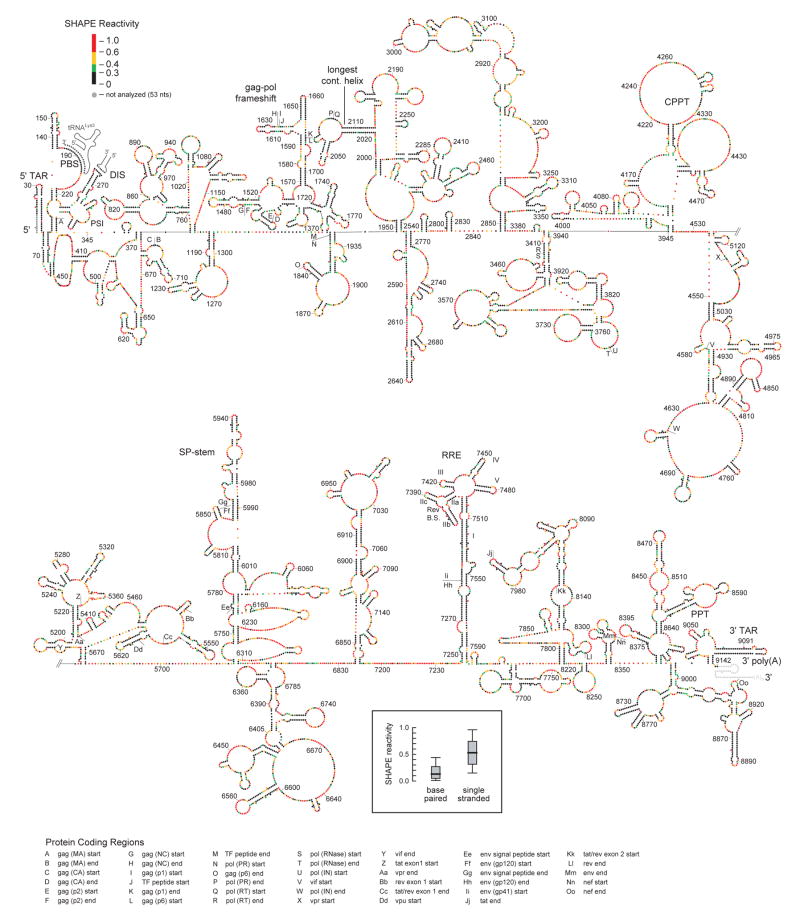
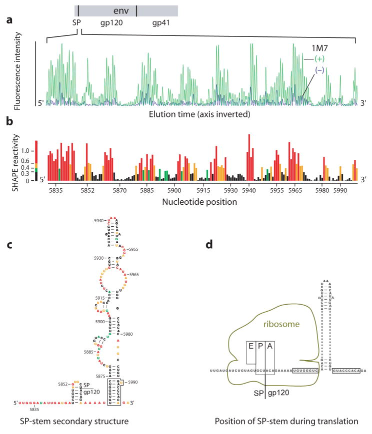
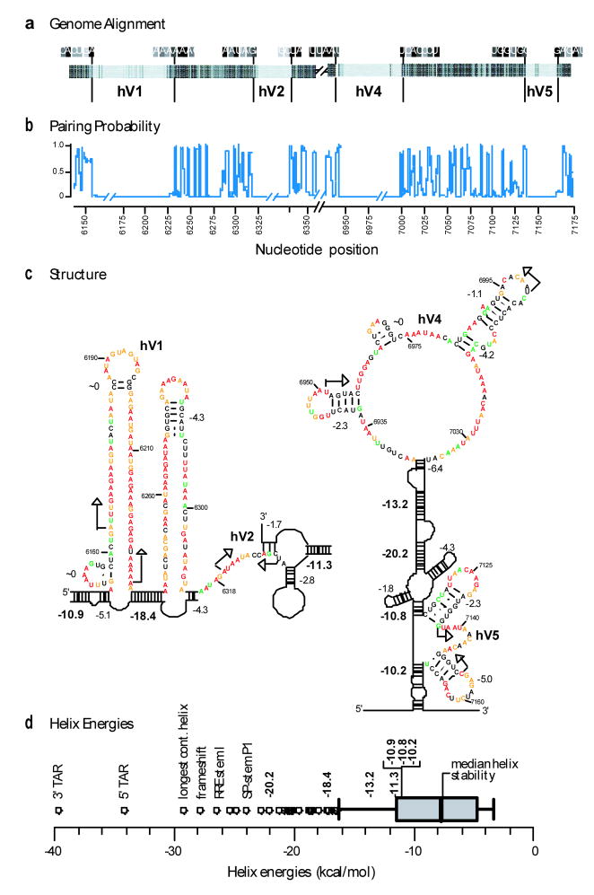
Single-stranded RNA viruses encompass broad classes of infectious agents and cause the common cold, cancer, AIDS and other serious health threats. Viral replication is regulated at many levels, including the use of conserved genomic RNA structures. Most potential regulatory elements in viral RNA genomes are uncharacterized. Here we report the structure of an entire HIV-1 genome at single nucleotide resolution using SHAPE, a high-throughput RNA analysis technology. The genome encodes protein structure at two levels. In addition to the correspondence between RNA and protein primary sequences, a correlation exists between high levels of RNA structure and sequences that encode inter-domain loops in HIV proteins. This correlation suggests that RNA structure modulates ribosome elongation to promote native protein folding. Some simple genome elements previously shown to be important, including the ribosomal gag-pol frameshift stem-loop, are components of larger RNA motifs. We also identify organizational principles for unstructured RNA regions, including splice site acceptors and hypervariable regions. These results emphasize that the HIV-1 genome and, potentially, many coding RNAs are punctuated by previously unrecognized regulatory motifs and that extensive RNA structure constitutes an important component of the genetic code.
Figures
Comment in
-
Structural biology: Aerial view of the HIV genome.Nature. 2009 Aug 6;460(7256):696-8. doi: 10.1038/460696a. Nature. 2009. PMID: 19661906 No abstract available.
References
-
- Cann AJ. Principles of Molecular Virology. Elsevier Academic Press; Amsterdam: 2005.
-
- Coffin JM, Hughes SH, Varmus HE. Retroviruses. Cold Spring Harbor Laboratory Press; Cold Spring Harbor, NY: 1997. - PubMed
-
- Frankel AD, Young JA. HIV-1: Fifteen proteins and an RNA. Annu Rev Biochem. 1998;67:1–25. - PubMed
-
- Damgaard CK, Andersen ES, Knudsen B, Gorodkin J, Kjems J. RNA interactions in the 5′ region of the HIV-1 genome. J Mol Biol. 2004;336:369–379. - PubMed
-
- Goff SP. Host factors exploited by retroviruses. Nature Rev Microbiol. 2007;5:253–263. - PubMed
Publication types
MeSH terms
Substances
Grants and funding
- R01 AI068462/AI/NIAID NIH HHS/United States
- T32 AI07419/AI/NIAID NIH HHS/United States
- R01 GM064803/GM/NIGMS NIH HHS/United States
- N01 CO012400/CA/NCI NIH HHS/United States
- AI44667/AI/NIAID NIH HHS/United States
- N01 CO012400/CO/NCI NIH HHS/United States
- R01 AI044667/AI/NIAID NIH HHS/United States
- HHSN261200800001C/RC/CCR NIH HHS/United States
- R56 AI044667/AI/NIAID NIH HHS/United States
- T32 AI007419/AI/NIAID NIH HHS/United States
- R37 AI044667/AI/NIAID NIH HHS/United States
- AI068462/AI/NIAID NIH HHS/United States
- N01-CO-12400/CO/NCI NIH HHS/United States
- HHSN261200800001E/CA/NCI NIH HHS/United States
LinkOut - more resources
Full Text Sources
Other Literature Sources
