. 2009 Oct 7;28(19):2992-3004.
doi: 10.1038/emboj.2009.226. Epub 2009 Aug 6.

A key role for Ctf4 in coupling the MCM2-7 helicase to DNA polymerase alpha within the eukaryotic replisome

Affiliations

A key role for Ctf4 in coupling the MCM2-7 helicase to DNA polymerase alpha within the eukaryotic replisome

Agnieszka Gambus et al. EMBO J. .

Abstract

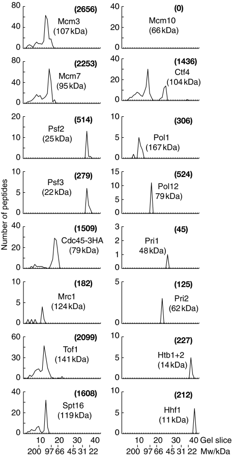
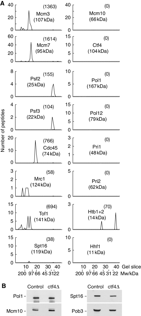
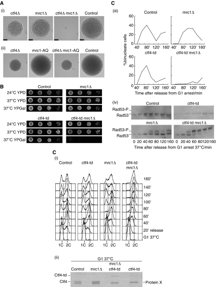
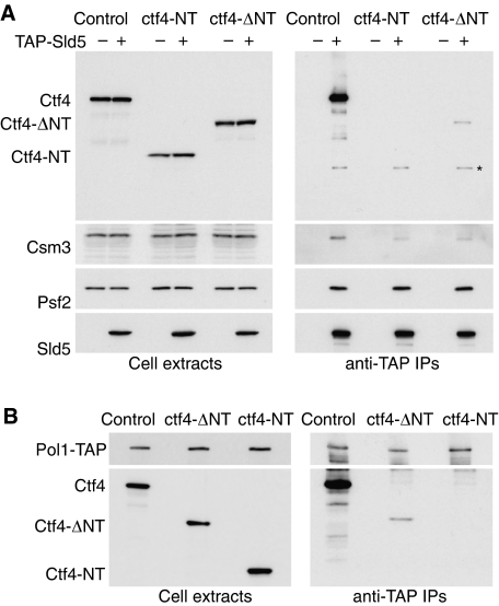
The eukaryotic replisome is a crucial determinant of genome stability, but its structure is still poorly understood. We found previously that many regulatory proteins assemble around the MCM2-7 helicase at yeast replication forks to form the replisome progression complex (RPC), which might link MCM2-7 to other replisome components. Here, we show that the RPC associates with DNA polymerase alpha that primes each Okazaki fragment during lagging strand synthesis. Our data indicate that a complex of the GINS and Ctf4 components of the RPC is crucial to couple MCM2-7 to DNA polymerase alpha. Others have found recently that the Mrc1 subunit of RPCs binds DNA polymerase epsilon, which synthesises the leading strand at DNA replication forks. We show that cells lacking both Ctf4 and Mrc1 experience chronic activation of the DNA damage checkpoint during chromosome replication and do not complete the cell cycle. These findings indicate that coupling MCM2-7 to replicative polymerases is an important feature of the regulation of chromosome replication in eukaryotes, and highlight a key role for Ctf4 in this process.


Conflict of interest statement

The authors declare that they have no conflict of interest.

Figures

Figure 1
Figure 1
The RPC associates with DNA polymerase α. A cell extract of TAP-SLD5 MCM4-5FLAG (YAG236-2) was generated in the presence of 50 mM potassium acetate and the RPC was purified as described in Materials and methods. The final material was separated in a 4–12% SDS–PAGE gradient gel and the lane was then cut into 40 slices. The graphs illustrate the total number of peptides that were identified by mass spectrometry in each slice of the gel, for a representative selection of the identified proteins. More details are shown in Supplementary Figure 1. The numbers in brackets indicate the combined Mascot score for each protein, corresponding to the sum of the unique peptide scores.
Figure 2
Figure 2
MCM2-7 associates with DNA polymerase α only during S-phase when RPCs are present. (A) An asynchronous culture of MCM4-5FLAG POL1-6HA PRI1-9MYC (YFJD62) was arrested in G1-phase at 24°C with mating pheromone before release into S-phase for the indicated times. DNA content was measured by flow cytometry. (B) Cell extracts were generated from the same experiment and treated with benzonase to digest chromosomal DNA as described in Materials and methods, before centrifugation and isolation of Mcm4-5FLAG by immunoprecipitation. The indicated proteins were analysed by immunoblotting. (C) Digestion of chromosomal DNA was monitored in an analogous experiment to that described in (A). Most DNA was digested during the initial 30′ treatment with Benzonase before centrifugation (compare lanes 1 and 2). The remainder was removed by centrifugation at 16 000 g for 30′ (lane 3). Even without centrifugation, all chromosomal DNA was degraded to undetectable levels during the time taken for the complete immunoprecipitation procedure (lane 4). (D) The experiment in (A) was repeated and samples taken 20′ after release from G1-phase. Benzonase and ethidium bromide were added to the initial extract as indicated, before immunoprecipitation of Mcm4.
Figure 3
Figure 3
Ctf4 is required for stable association of the RPC with DNA polymerase α. (A) RPC material was purified from ctf4Δ TAP-SLD5 MCM4-5FLAG (YAG374-2) and analysed as described above for Figure 1. The graphs illustrate a representative selection of the identified proteins, and the full data set is shown in Supplementary Figure 2. (B) The levels of Pol1, Mcm10, Spt16 and Pob3 were monitored by immunoblotting in extracts of control and ctf4Δ cells.
Figure 4
Figure 4
A fraction of Ctf4 can interact with GINS throughout the cell cycle and not just in RPCs. (A) Cultures of PSF2-TAP (YAG187-1) were grown at 24°C either asynchronously (Asyn.), or arrested in G1-phase with mating pheromone (G1), or released into S-phase from G1 in the presence of 0.2 M hydroxyurea for 60′ (S), or arrested in G2/M-phase with nocodazole (G2/M). Psf2-TAP was isolated by immunoprecipitation and the indicated proteins analysed by immunoblotting. (B) A cdc6Δ∷GAL-CDC6 TAP-SLD5 strain (YAG258-1) was grown at 24°C in rich medium containing galactose (YPGal), in parallel with a TAP-SLD5 control strain (YAG236-2), and cells were arrested in G2-M-phase with nocodazole. Expression of GAL-CDC6 was then repressed in fresh medium containing glucose (YPD) as well as nocodazole, before cells were released into fresh YPD medium to allow synchronization in the following G1-phase with mating pheromone. At this stage, the MCM helicase was assembled at origins into prereplicative complexes in the control strain (+ pre-RCs), but not in the GAL-CDC6 strain (− pre-RCs). Finally, cells were released into S-phase for 60′ in the presence of 0.2 M HU. TAP-Sld5 was isolated by immunoprecipitation and the indicated proteins analysed by immunoblotting.
Figure 5
Figure 5
GINS and Ctf4 interact directly to form a stable complex. (A) Recombinant GINS was purified from an E. coli cell extract that also contained recombinant Ctf4 as described in Materials and methods, by virtue of a GST tag on the Psf3 subunit of GINS (E1). The purified material was separated in an SDS–PAGE gel that was stained with Coomassie blue. The band marked by an asterisk (*) was identified by mass spectrometry and corresponds to free GST that might have been derived as a degradation product from GST-Psf3. Ctf4 was then isolated from the purified GINS sample using a six-histidine tag at its amino terminus, and the figure shows the flow through of this purification step (FT), the washes (W1–W3), and the final eluate that represents purified GINS–Ctf4 complex (E2). The identity of the various bands was confirmed by mass spectrometry. (B) A similar experiment was performed using an E. coli extract containing recombinant Sld5–Psf2 subcomplex of GINS (with the GST tag on Sld5), together with 6His-Ctf4. (C) Summary of the interaction of truncated forms of recombinant Ctf4 with GINS in an E. coli extract, in an assay analogous to that described above. (D) An extract of E. coli was generated that contained recombinant Ctf4-ΔNT with 6His at its amino terminus, and recombinant GINS with the Streptag III epitope at the amino terminus of Psf3 as well as a truncated form of Psf1 (Psf1-ΔCT, amino acids 1–164) to improve visualisation of the four GINS proteins in the subsequent gel. StreptagIII-Psf3 was isolated from the extract and 6His-Ctf4ΔNT was then purified from the resultant material. The purified complex of GINS and Ctf4ΔNT was applied to a gel filtration column, and migrated with a retention volume of 50 ml (distinct from the void volume of 46 ml). The peak fractions were combined and concentrated before separation in an SDS–PAGE gel that was then stained with Coomassie blue. (E) Comparison of the migration through gel filtration columns of purified recombinant versions of GINS-Ctf4ΔNT, GINS and Ctf4ΔNT.
Figure 6
Figure 6
Direct interaction of Ctf4 with the amino terminus of Pol1 is independent of the WD40 domain of Ctf4. (A) Summary of fragments of Pol1 that were isolated in a two-hybrid screen with amino acids 461–927 of Ctf4 (Ctf4-ΔNT) as bait. (B) An extract of E. coli was generated that contained the indicated recombinant versions of Ctf4, each with 6His at the amino terminus, together with Pol1NT-Myc (left panel). The 6His-Ctf4 fragments were isolated from the extract (right panel) and the presence of Pol1NT-Myc and Ctf4 determined by immunoblotting.
Figure 7
Figure 7
Cells lacking both Ctf4 and Mrc1 undergo chronic checkpoint activation during chromosome replication and cannot complete the cell cycle. (A) (i) A ctf4Δ strain was crossed to mrc1Δ and the subsequent diploid was sporulated and subjected to tetrad analysis. Cells were grown at 30°C for 24 h and then photographed, and the images show a complete tetrad that illustrates the growth of control and mutant cells. The scale-bar corresponds to 50 μm. (ii) Similar analysis of a cross of ctf4Δ to mrc1-AQ. (B) Serial dilutions of the indicated strains were placed on the indicated media (YPD, yeast extract, peptone, dextrose; YPGal, yeast extract, peptone, galactose) and cells were grown for 48 h before the images were taken. (C) Asynchronous cultures of control (YKL200), mrc1Δ (YBH81), ctf4-td (YAG138-1) and ctf4-td mrc1Δ (YAG156-1) were grown at 24°C in YPRaffinose medium, and then synchronized in G1-phase with mating pheromone. Cells were transferred to YPGalactose medium for 35 min at 24°C to induce expression of the Ubr1 E3 ubiquitin ligase, and then shifted to 37°C for 1 h to inactivate Ctf4-td. Cells were transferred subsequently to fresh medium lacking mating pheromone to allow them to proceed with the cell cycle, and samples were taken at the indicated times and processed for flow cytometry (i), immunoblotting to monitor Ctf4—Protein X indicates a non-specific band in the immunoblot (ii), fluorescence microscopy to analyse nuclear division (iii), or used to generate protein extracts to monitor phosphorylation of the Rad53 checkpoint kinase. Rad53-P indicates the hyperphosphorylated form of Rad53 (iv).
Figure 8
Figure 8
The WD40 domain of Ctf4 is not essential for interaction of Ctf4 with GINS and DNA polymerase α in vivo. (A) Cell extracts were generated from asynchronous cultures of the indicated strains, and TAP-Sld5 isolated by immunoprecipitation with IgG-Sepharose, before detection of the indicated proteins by immunoblotting. The asterisk in the Ctf4 immunoblot of the immunoprecipitates indicates TAP-Sld5, which binds in a non-specific manner to antibodies by virtue of the TAP tag. (B) A similar experiment was performed with asynchronous cultures of the indicated strains expressing Pol1-TAP.
Figure 9
Figure 9
The ability of Ctf4 to bind GINS and DNA polymerase α is uniquely important in cells lacking Mrc1. (A) Spores of the indicated genotypes were identified by tetrad analysis and grown for 24 h at 30°C before imaging. The scale-bar corresponds to 50 μm. (B) Asynchronous cultures of the indicated strains were grown at 30°C and images were taken by phase contrast microscopy (left panels)—the scale-bar corresponds to 5 μm. Samples were also processed for flow cytometry (right panels). (C) (i) Cells were grown on the indicated medium for 48 h at 30°C before the images were taken. (ii) Strains lacking Ctf4 or with the indicated truncated versions of Ctf4 were crossed to a ctf18Δ strain and subjected to tetrad analysis. Images of the indicated double mutants are shown after 24 h growth at 30°C. (iii) Strains with the same range of ctf4 mutations were crossed to an mrc1Δ strain and processed as above.
Figure 10
Figure 10
Ctf4 couples DNA polymerase α to the RPC. Ctf4 binds directly to DNA polymerase α and to GINS in budding yeast, and is required to link DNA polymerase α to the RPC at replication forks. Mrc1 is a strong candidate for a factor that might couple DNA polymerase epsilon to the RPC, although this remains to be shown directly. The eukaryotic replisome will contain other components in addition to those depicted, but the interaction of such factors with the RPC and with DNA polymerases remains to be characterized in the future.

Similar articles

Cited by

References

    1. Aguilera A, Gomez-Gonzalez B (2008) Genome instability: a mechanistic view of its causes and consequences. Nat Rev Genet 9: 204–217 - PubMed
    1. Alcasabas AA, Osborn AJ, Bachant J, Hu F, Werler PJ, Bousset K, Furuya K, Diffley JF, Carr AM, Elledge SJ (2001) Mrc1 transduces signals of DNA replication stress to activate Rad53. Nat Cell Biol 3: 958–965 - PubMed
    1. Alvaro D, Lisby M, Rothstein R (2007) Genome-wide analysis of Rad52 foci reveals diverse mechanisms impacting recombination. PLoS Genet 3: e228. - PMC - PubMed
    1. Aparicio OM, Weinstein DM, Bell SP (1997) Components and dynamics of DNA replication complexes in S. cerevisiae: redistribution of MCM complexes and Cdc45p during S phase. Cell 91: 59–69 - PubMed
    1. Aparicio T, Guillou E, Coloma J, Montoya G, Mendez J (2009) The human GINS complex associates with Cdc45 and MCM and is essential for DNA replication. Nucleic Acids Res 37: 2087–2095 - PMC - PubMed

Publication types

MeSH terms