

Recognition of an intra-chain tandem 14-3-3 binding site within PKCepsilon
- PMID: 19662078
- PMCID: PMC2750047
- DOI: 10.1038/embor.2009.150
Recognition of an intra-chain tandem 14-3-3 binding site within PKCepsilon
Abstract
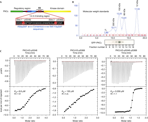
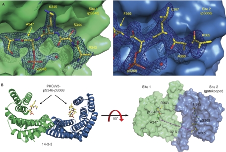
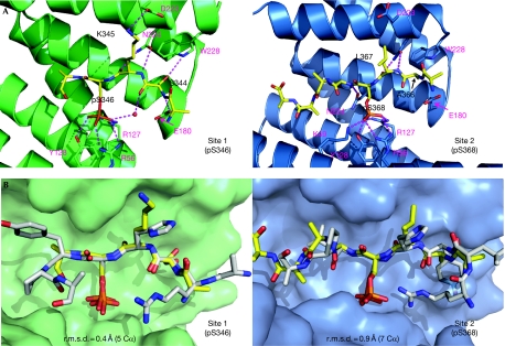
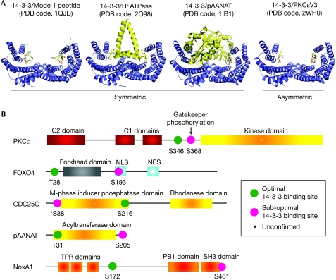
The phosphoserine/threonine binding protein 14-3-3 stimulates the catalytic activity of protein kinase C-epsilon (PKCepsilon) by engaging two tandem phosphoserine-containing motifs located between the PKCepsilon regulatory and catalytic domains (V3 region). Interaction between 14-3-3 and this region of PKCepsilon is essential for the completion of cytokinesis. Here, we report the crystal structure of 14-3-3zeta bound to a synthetic diphosphorylated PKCepsilon V3 region revealing how a consensus 14-3-3 site and a divergent 14-3-3 site cooperate to bind to 14-3-3 and so activate PKCepsilon. Thermodynamic data show a markedly enhanced binding affinity for two-site phosphopeptides over single-site 14-3-3 binding motifs and identifies Ser 368 as a gatekeeper phosphorylation site in this physiologically relevant 14-3-3 ligand. This dual-site intra-chain recognition has implications for other 14-3-3 targets, which seem to have only a single 14-3-3 motif, as other lower affinity and cryptic 14-3-3 gatekeeper sites might exist.
Conflict of interest statement
The authors declare that they have no conflict of interest.
Figures
Similar articles
-
The identification and characterization of novel PKCepsilon phosphorylation sites provide evidence for functional cross-talk within the PKC superfamily.Biochem J. 2008 Apr 15;411(2):319-31. doi: 10.1042/bj20071348. Biochem J. 2008. PMID: 18237277
-
PKCepsilon increases phosphorylation of the cardiac myosin binding protein C at serine 302 both in vitro and in vivo.Biochemistry. 2007 Jun 12;46(23):7054-61. doi: 10.1021/bi700467k. Epub 2007 May 16. Biochemistry. 2007. PMID: 17503784 Free PMC article.
-
The regulated assembly of a PKCepsilon complex controls the completion of cytokinesis.Nat Cell Biol. 2008 Aug;10(8):891-901. doi: 10.1038/ncb1749. Epub 2008 Jul 6. Nat Cell Biol. 2008. PMID: 18604201
-
Structural determinants of 14-3-3 binding specificities and regulation of subcellular localization of 14-3-3-ligand complexes: a comparison of the X-ray crystal structures of all human 14-3-3 isoforms.Semin Cancer Biol. 2006 Jun;16(3):173-82. doi: 10.1016/j.semcancer.2006.03.007. Epub 2006 Apr 1. Semin Cancer Biol. 2006. PMID: 16678437 Review.
-
Does isoform diversity explain functional differences in the 14-3-3 protein family?Curr Pharm Biotechnol. 2006 Jun;7(3):217-23. doi: 10.2174/138920106777549777. Curr Pharm Biotechnol. 2006. PMID: 16789906 Review.
Cited by
-
A Supramolecular Stabilizer of the 14-3-3ζ/ERα Protein-Protein Interaction with a Synergistic Mode of Action.Angew Chem Int Ed Engl. 2020 Mar 23;59(13):5284-5287. doi: 10.1002/anie.201914517. Epub 2020 Feb 11. Angew Chem Int Ed Engl. 2020. PMID: 31814236 Free PMC article.
-
Mechanism of IRSp53 inhibition by 14-3-3.Nat Commun. 2019 Jan 29;10(1):483. doi: 10.1038/s41467-019-08317-8. Nat Commun. 2019. PMID: 30696821 Free PMC article.
-
Concatenation of 14-3-3 with partner phosphoproteins as a tool to study their interaction.Sci Rep. 2019 Oct 18;9(1):15007. doi: 10.1038/s41598-019-50941-3. Sci Rep. 2019. PMID: 31628352 Free PMC article.
-
Phosphorylation dependence and stoichiometry of the complex formed by tyrosine hydroxylase and 14-3-3γ.Mol Cell Proteomics. 2014 Aug;13(8):2017-30. doi: 10.1074/mcp.M113.035709. Epub 2014 Jun 19. Mol Cell Proteomics. 2014. PMID: 24947669 Free PMC article.
-
Co-ordinated control of the Aurora B abscission checkpoint by PKCε complex assembly, midbody recruitment and retention.Biochem J. 2021 Jun 25;478(12):2247-2263. doi: 10.1042/BCJ20210283. Biochem J. 2021. PMID: 34143863 Free PMC article.
References
-
- Adams PD, Grosse-Kunstleve RW, Hung LW, Ioerger TR, McCoy AJ, Moriarty NW, Read RJ, Sacchettini JC, Sauter NK, Terwilliger TC (2002) PHENIX: building new software for automated crystallographic structure determination. Acta Crystallogr D Biol Crystallogr 58: 1948–1954 - PubMed
-
- Bridges D, Moorhead GB (2005) 14-3-3 proteins: a number of functions for a numbered protein. Sci STKE 2005: re10. - PubMed
-
- Delano WL (2002) The PyMOL Molecular Graphics System. Palo Alto, CA USA: Delano Scientific. http://www.pymol.org
-
- Durgan J, Cameron AJ, Saurin AT, Hanrahan S, Totty N, Messing RO, Parker PJ (2008) The identification and characterization of novel PKCepsilon phosphorylation sites provide evidence for functional cross-talk within the PKC superfamily. Biochem J 411: 319–331 - PubMed
-
- Emsley P, Cowtan K (2004) Coot: model-building tools for molecular graphics. Acta Crystallogr D Biol Crystallogr 60: 2126–2132 - PubMed
Publication types
MeSH terms
Substances
Grants and funding
LinkOut - more resources
Full Text Sources
Molecular Biology Databases
