. 2009 Aug;5(8):e1000542.
doi: 10.1371/journal.ppat.1000542. Epub 2009 Aug 7.

Anopheles gambiae PGRPLC-mediated defense against bacteria modulates infections with malaria parasites

Affiliations

Anopheles gambiae PGRPLC-mediated defense against bacteria modulates infections with malaria parasites

Stephan Meister et al. PLoS Pathog. 2009 Aug.

Abstract

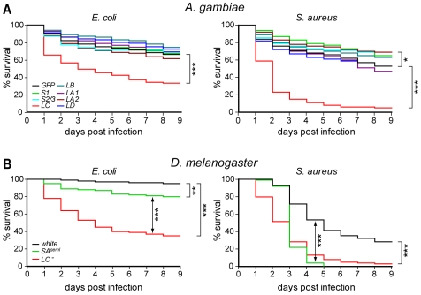
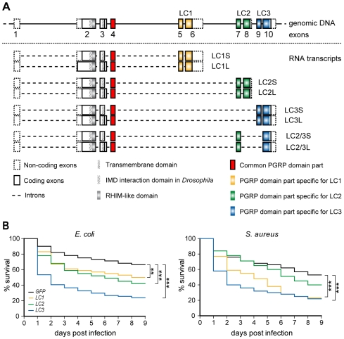
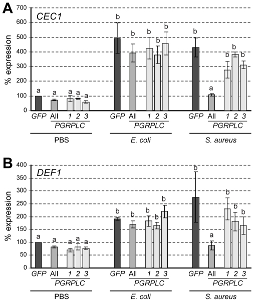
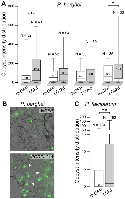
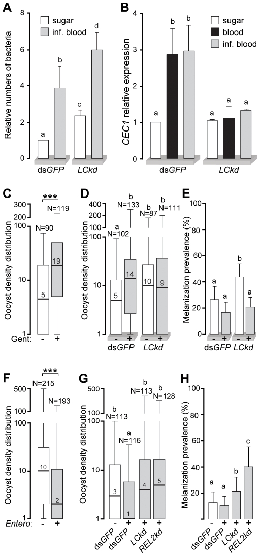
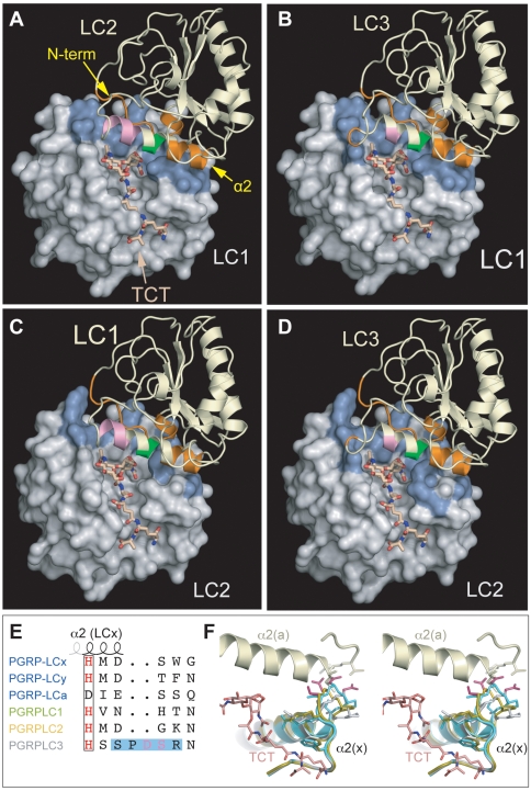
Recognition of peptidoglycan (PGN) is paramount for insect antibacterial defenses. In the fruit fly Drosophila melanogaster, the transmembrane PGN Recognition Protein LC (PGRP-LC) is a receptor of the Imd signaling pathway that is activated after infection with bacteria, mainly Gram-negative (Gram-). Here we demonstrate that bacterial infections of the malaria mosquito Anopheles gambiae are sensed by the orthologous PGRPLC protein which then activates a signaling pathway that involves the Rel/NF-kappaB transcription factor REL2. PGRPLC signaling leads to transcriptional induction of antimicrobial peptides at early stages of hemolymph infections with the Gram-positive (Gram+) bacterium Staphylococcus aureus, but a different signaling pathway might be used in infections with the Gram- bacterium Escherichia coli. The size of mosquito symbiotic bacteria populations and their dramatic proliferation after a bloodmeal, as well as intestinal bacterial infections, are also controlled by PGRPLC signaling. We show that this defense response modulates mosquito infection intensities with malaria parasites, both the rodent model parasite, Plasmodium berghei, and field isolates of the human parasite, Plasmodium falciparum. We propose that the tripartite interaction between mosquito microbial communities, PGRPLC-mediated antibacterial defense and infections with Plasmodium can be exploited in future interventions aiming to control malaria transmission. Molecular analysis and structural modeling provided mechanistic insights for the function of PGRPLC. Alternative splicing of PGRPLC transcripts produces three main isoforms, of which PGRPLC3 appears to have a key role in the resistance to bacteria and modulation of Plasmodium infections. Structural modeling indicates that PGRPLC3 is capable of binding monomeric PGN muropeptides but unable to initiate dimerization with other isoforms. A dual role of this isoform is hypothesized: it sequesters monomeric PGN dampening weak signals and locks other PGRPLC isoforms in binary immunostimulatory complexes further enhancing strong signals.


Conflict of interest statement

The authors have declared that no competing interests exist.

Figures

Figure 1
Figure 1. Resistance of PGRP kd mosquitoes and mutant fruit flies to bacterial infections.
(A) Kaplan Meier survival curves of adult A. gambiae females silenced for PGRP gene expression, and infected 4 days later with E. coli (left) or S. aureus (right). PGRPS2 and S3 were concomitantly silenced using dsRNA that fully matched both sequences. Survival was recorded daily for 9 days and compared to that of GFP dsRNA-injected controls. The data are the average of very similar results obtained from at least three replicate infections. (B) Kaplan Meier survival curves of adult D. melanogaster females of the PGRP-SAseml, PGRP-LC and white (control) mutant strains infected with E. coli (left) or S. aureus (right).
Figure 2
Figure 2. Contribution of A. gambiae PGRPLC isoforms generated through alterative splicing in antibacterial defense.
(A) Genomic organization of the PGRPLC locus and alternative splice variants identified through genomic PCR and RT-PCR reactions. Sequences encoding variable parts of the 3 PGRP domains and the sequence encoding the common part of all PGRP domains are depicted in different colors. (B) Percent survival rates of A. gambiae females silenced for the expression of each of the 3 main PGRPLC isoforms or all isoforms simultaneously and infected 4 days later with E. coli (left) or S. aureus (right). Survival was recorded daily, for 5 days. GFP dsRNA-injected controls were used as controls. Colors corresponding to those used to indicate different isoforms in (A). Error bars represent standard errors of three or more replicate infections.
Figure 3
Figure 3. Role of PGRPLC in AMP expression following bacterial infections.
Relative percent abundance of CEC1 (A) and DEF1 (B) transcripts in PGRPLC kd and dsGFP-treated control A. gambiae adult females, 3 h after injection with saline solution (PBS), E. coli or S. aureus. Error bars indicate standard deviations. Same letters above each bar represent statistically similar expression values while different letters indicate statistically significant differences (P<0.001) as determined by multiple comparisons using the Student's t-test.
Figure 4
Figure 4. PGRPLC affects mosquito infections with Plasmodium.
(A) Box plots of median numbers and distribution of oocyst intensities in P. berghei-infected dsGFP-treated (controls) or PGRPLC kd A. gambiae females. Independent controls were used for each of the entire PGRPLC gene (LC) and isoform-specific kds. Boxes show the distribution of 50% of the data and whiskers indicate the full range. N above each whisker indicates the numbers of mosquitoes. Results of Mann Whitney statistical tests are shown above each box plot: ***, P<0.001; *, P<0.05. (B) P. berghei 7-day-old oocysts and melanized ookinetes (arrowheads) in the midgut of PGRPLC kd and dsGFP-treated control A. gambiae females. (C) Box plots of median numbers and distribution of oocyst intensities of P. falciparum field isolates in dsLacZ-treated control or PGRPLC kd A. gambiae. **, P<0.01.
Figure 5
Figure 5. PGRPLC controls gut bacteria modulating Plasmodium infections.
(A) Relative numbers of bacteria in dsGFP-treated control or PGRPLC kd A. gambiae females, fed on sugar or P. berghei-infected mice 24 h before sampling. Quantification was performed by quantitative genomic PCR of a conserved bacterial 16S rDNA fragment and referenced to sugar-fed controls. Error bars indicate standard errors. Comparisons between all the samples were performed using the Student's t-test and the results are shown as letters above each bar. Same letters indicate no significant difference, while different letters indicate at least P<0.05. In this graph all P values were <0.001. (B) CEC1 relative expression in dsGFP-treated control and PGRPLC kd A. gambiae females fed on sugar, naïve blood or P. berghei-infected blood. Expression in sugar-fed controls is used as a reference. As in (A) different letters above each bar indicate Student's t-test P value<0.001. (C) Median numbers and distribution of P. berghei oocyst intensities in Gentamycin-treated (+) and untreated (−) mosquitoes. Boxes include 50% of the data and whiskers indicate the range in a log10-transformed scale. Median is shown with the bar and number within each box. ***, P<0.001 of Mann Whitney test. (D) Median numbers and distribution of P. berghei oocyst intensities in Gentamycin-treated (+) and untreated (−) PGRPLC kd and dsGFP-treated controls shown in a log10-transformed scale. As above, different letters above each dataset indicate statistically significant differences: P<0.001 for dsGFP(−)/dsGFP(+) and P<0.01 for dsGFP(−)/LCkd(−) and dsGFP(−)/LCkd(+). (E) Prevalence of melanized ookinetes in the mosquitoes presented in (D). Statistical analysis was performed with the Fisher's exact test: dsGFP(−)/LCkd(−), P<0.05; dsGFP(+)/LCkd(−), P<0.0001; LCkd(−)/LCkd(+), P<0.001. (F) Median numbers and distribution of P. berghei oocyst intensities in Enterobacter-infected (+) and non-infected (−) mosquitoes. Note that the y-axis is log10-transformed. (G) Median numbers and distribution of P. berghei oocyst intensities in Enterobacter-infected (+) and non-infected (−) dsGFP-treated mosquitoes, and in Enterobacter-infected PGRPLC kd and REL2 kd mosquitoes. Different letters above each dataset indicate statistically significant differences as follows: P<0.005 for dsGFP(−)/dsGFP(+) and P<0.001 for dsGFP(+)/LCkd(+) and dsGFP(−)/LCkd(+). (H) Prevalence of melanized ookinetes in the mosquitoes presented in (G). dsGFP(−)/LCkd(+), P<0.05; dsGFP(−)/REL2kd(−), P<0.0001; dsGFP(+)/LCkd(+), P<0.0001; dsGFP(+)/REL2kd(+), P<0.05; LCkd(+)/REL2kd(+), P<0.005.
Figure 6
Figure 6. TCT-mediated hetero-dimerization of Anopheles PGRPLC isoforms.
Heterodimer models of AgPGRPLC1-TCT-LC2 (A), AgPGRPLC1-TCT-LC3 (B), AgPGRPLC2-TCT-LC1 (C) and AgPGRPLC2-TCT-LC3 (D). PGRPLC/x molecules are shown in molecular surface models and PGRPLC/a in ribbon diagrams. The PGRPLC/a N-terminus and helix α2 that mediate dimerization are indicated, with monomer-interacting parts colored in orange, parts contacting both monomer and TCT in green and the TCT-interacting part in pink. Interface residues on the surface of PGRPLC/x are shown in blue. (E) Detail alignment of the PD-loop between Ag and Dm PGRPLCs, highlighting the modeled loop and clashing residues. (F) Stereo view of the putative dimer interface at the contact between helix α2/PD-loop of AgPGRPLC3/x and helix α2 of AgPGRPLC1/a (pale green). Three alternative AgPGRPLC3/x models corresponding to different PD-loop modeling approaches are superimposed; in grey the model from MODELLER, in gold the average model structure from ARIA; and in turquoise the Robetta model. PD-loop Residues D61 and S62 (magenta), which clash severely with helix a2 in the three models, and the anchor, TCT-interacting residues R63 and F65, are shown in sticks.

References

    1. Michel T, Reichhart JM, Hoffmann JA, Royet J. Drosophila Toll is activated by Gram-positive bacteria through a circulating peptidoglycan recognition protein. Nature. 2001;414:756–759. - PubMed
    1. Bischoff V, Vignal C, Boneca IG, Michel T, Hoffmann JA, et al. Function of the drosophila pattern-recognition receptor PGRP-SD in the detection of Gram-positive bacteria. Nat Immunol. 2004;5:1175–1180. - PubMed
    1. Gottar M, Gobert V, Michel T, Belvin M, Duyk G, et al. The Drosophila immune response against Gram-negative bacteria is mediated by a peptidoglycan recognition protein. Nature. 2002;416:640–644. - PubMed
    1. Choe KM, Werner T, Stoven S, Hultmark D, Anderson KV. Requirement for a peptidoglycan recognition protein (PGRP) in Relish activation and antibacterial immune responses in Drosophila. Science. 2002;296:359–362. - PubMed
    1. Kaneko T, Yano T, Aggarwal K, Lim JH, Ueda K, et al. PGRP-LC and PGRP-LE have essential yet distinct functions in the drosophila immune response to monomeric DAP-type peptidoglycan. Nat Immunol. 2006;7:715–723. - PubMed

Publication types

MeSH terms