

Age-related differences in multiple measures of white matter integrity: A diffusion tensor imaging study of healthy aging
- PMID: 19662658
- PMCID: PMC2826569
- DOI: 10.1002/hbm.20872
Age-related differences in multiple measures of white matter integrity: A diffusion tensor imaging study of healthy aging
Abstract
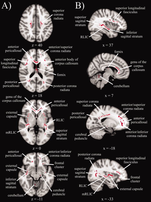
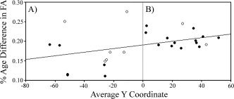
Diffusion tensor imaging (DTI) measures diffusion of molecular water, which can be used to calculate indices of white matter integrity. Early DTI studies of aging primarily focused on two global measures of integrity; the average rate (mean diffusivity, MD) and orientation coherence (fractional anisotropy, FA) of diffusion. More recent studies have added measures of water movement parallel (axial diffusivity, AD) and perpendicular (radial diffusivity, RD) to the primary diffusion direction, which are thought to reflect the neural bases of age differences in diffusion (i.e., axonal shrinkage and demyelination, respectively). In this study, patterns of age differences in white matter integrity were assessed by comparing younger and healthy older adults on multiple measures of integrity (FA, AD, and RD). Results revealed two commonly reported patterns (Radial Increase Only and Radial/Axial Increase), and one relatively novel pattern (Radial Increase/Axial Decrease) that varied by brain region and may reflect differential aging of microstructural (e.g., degree of myelination) and macrostructural (e.g., coherence of fiber orientation) properties of white matter. In addition, larger age differences in FA in frontal white matter were consistent with the anterior-posterior gradient of age differences in white matter integrity. Together, these findings complement other recent studies in providing information about patterns of diffusivity that are characteristic of healthy aging.
2009 Wiley-Liss, Inc.
Figures
References
-
- Abe O, Aoki S, Hayashi N, Yamada H, Kunimatsu A, Mori H, Yoshikawa T, Okubo T, Ohtomo K ( 2002): Normal aging in the central nervous system: Quantitative MR diffusion‐tensor analysis. Neurobiol Aging 23: 433–441. - PubMed
-
- Aboitiz F, Scheibel AB, Fisher RS, Zaidel E ( 1992): Fiber composition of the human corpus callosum. Brain Res 598: 143–153. - PubMed
-
- Ardekani S, Kumar A, Bartzokis G, Sinha U ( 2007): Exploratory voxel‐based analysis of diffusion indices and hemispheric asymmetry in normal aging. J Magn Reson Imaging 25: 154–167. - PubMed
-
- Basser PJ, Pierpaoli C ( 1996): Microstructural and physiological features of tissues elucidated by quantitative‐diffusion‐tensor MRI. J Magn Reson B 111: 209–219. - PubMed
Publication types
MeSH terms
Grants and funding
LinkOut - more resources
Full Text Sources
Medical
Miscellaneous
