. 2009 Jun;118(6):435-41.
doi: 10.1177/000348940911800606.

Techniques of celloidin removal from temporal bone sections

Affiliations

Techniques of celloidin removal from temporal bone sections

Jennifer T O'Malley et al. Ann Otol Rhinol Laryngol. 2009 Jun.

Abstract

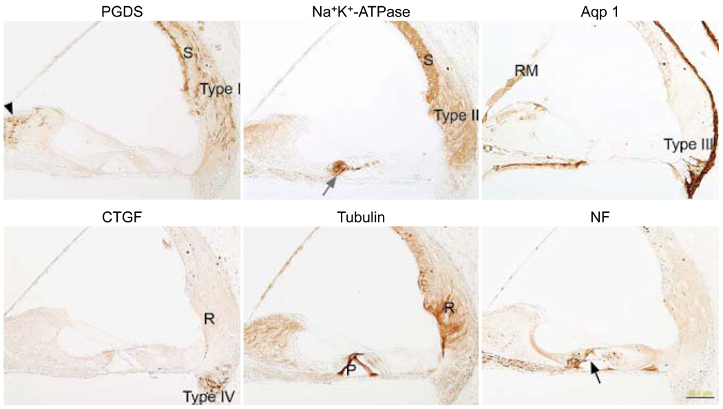
Objectives: We sought to determine whether the technique of celloidin removal influences the results of immunostaining in celloidin-embedded cochleae.

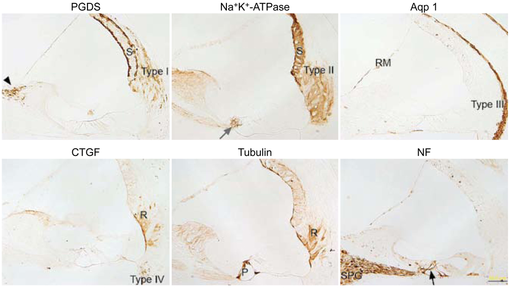
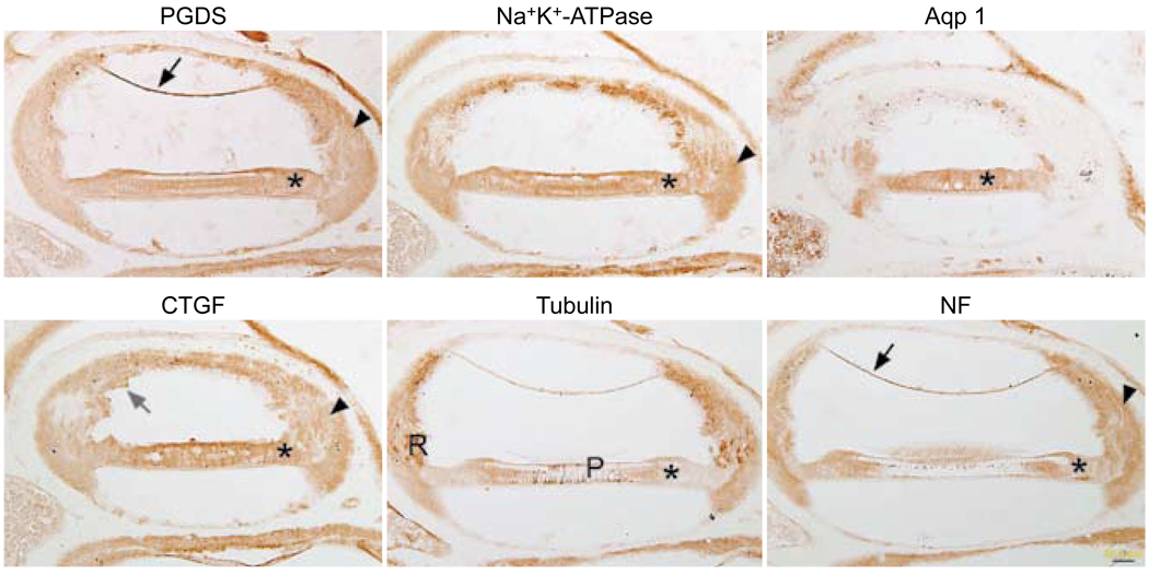
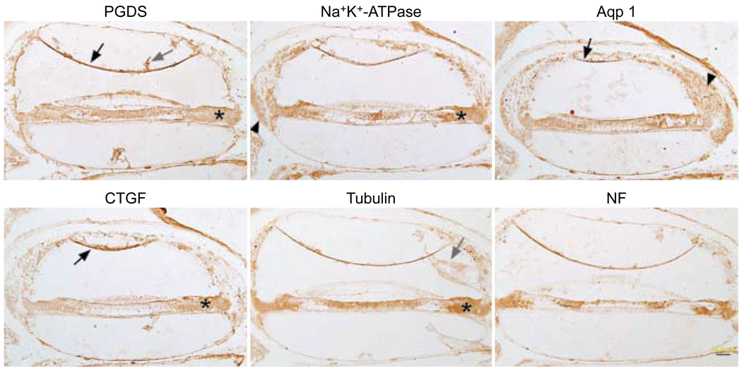
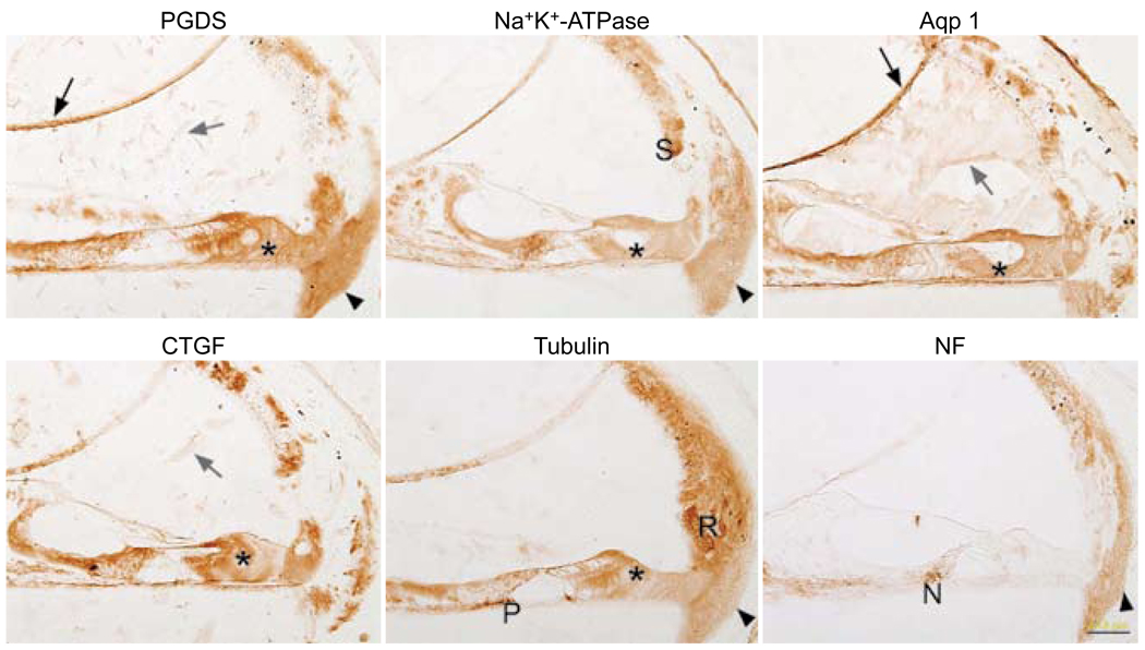
Methods: We compared four protocols of celloidin removal, including those using clove oil, acetone, ether-alcohol, and methanol saturated with sodium hydroxide. By optimally fixing our tissue (perfused mice), and keeping constant the fixative type (formalin plus acetic acid), fixation time (25 hours), and decalcification time (ethylenediaminetetraacetic acid for 7 days), we determined whether the technique of celloidin removal influenced the immunostaining results. Six antibodies were used with each removal method: prostaglandin D synthase, sodium, potassium adenosine triphosphatase (Na+,K(+)-ATPase), aquaporin 1, connective tissue growth factor, tubulin, and 200 kd neurofilament.

Results: Clove oil, acetone, and ether-alcohol resulted in incomplete removal of the celloidin, thereby negatively affecting the results of immunostaining. The methanol-sodium hydroxide method was effective in completely removing the celloidin; it produced the cleanest and most reproducible immunostaining for all six antibodies.

Conclusions: Freshly prepared methanol saturated with sodium hydroxide and diluted 1:2 with methanol was the best solvent for removing celloidin from mouse temporal bone sections, resulting in consistent and reproducible immunostaining with the six antibodies tested.


Figures

Fig. 1
Fig. 1
Paraffin sections immunostained with each of six antibodies. Prostaglandin D synthase (PGDS) antibody stains basal and marginal cells of stria vascularis (S), type I fibrocytes of spiral ligament (type I), and fibrocytes of spiral limbus (black arrowhead). Sodium, potassium adenosine triphosphatase (Na+,K+-ATPase) antibody stains stria vascularis (S), type II fibrocytes of spiral ligament (type II), and nerve fibers below inner hair cells (gray arrow). Antibody against aquaporin 1 (Aqp 1) stains type III fibrocytes of spiral ligament (type III) and medial portion of Reissner’s membrane (RM). Connective tissue growth factor (CTGF) antibody stains type IV fibrocytes of spiral ligament (type IV) and root cells (R). Antibody against tubulin stains pillar cells (P) and root cells (R). Neurofilament (NF) antibody stains some of spiral ganglion cells (SPG) and nerve fibers in Rosenthal’s canal, beneath inner and outer hair cells and crossing tunnel of Corti (black arrow). Calibration bar — 50 µm.
Fig. 2
Fig. 2
Celloidin removal with clove oil followed by immunostaining. All six panels show similar nonspecific staining pattern, regardless of antibody used. Reissner’s membrane (black arrows) shows background staining with PGDS and NF. Supporting cells (asterisks) show similar staining patterns with all six antibodies. Staining in spiral ligament (arrowheads) is diffusely brown with most of antibodies. In CTGF-stained tissue, bits of celloidin (gray arrow) are still evident in section near stria vascularis. Tubulin antibody starts to stain expected structures such as root cells (R) and pillar cells (P), but there is also great deal of brown background staining in section. Calibration bar — 50 µm.
Fig. 3
Fig. 3
Celloidin removal with ether-alcohol followed by immunostaining. All six panels show similar nonspecific staining pattern, regardless of antibody used. Once again, Reissner’s membrane (black arrows) stains with most of antibodies. Supporting cells (asterisks) are all diffusely brown. There is spurious staining in spiral ligament (arrowheads). Celloidin is still evident in many sections (gray arrows). Calibration bar — 50 µm.
Fig. 4
Fig. 4
Celloidin removal with acetone followed by immunostaining. Reissner’s membrane (black arrows) stains in many panels (nonspecific staining). Supporting cells (asterisks) are diffusely brown in many panels. With Na+,K+-ATPase antibody, stria (S) starts to stain appropriately, but there is a great deal of background staining. Celloidin (gray arrows) can still be seen in some panels. Spiral ligament (arrowheads) shows background staining. Tubulin panel shows appropriate staining in root cells (R) and in a portion of pillar cells (P), but background staining is also seen. Neurofilament antibody stains nerve fibers (N) below inner hair cell area, but it also spuriously stains spiral ligament (arrowhead). Calibration bar — 50 µm.
Fig. 5
Fig. 5
Celloidin removal with methanol saturated with sodium hydroxide followed by immunostaining. Successful immunostaining for all six antibodies is possible when methanol saturated with sodium hydroxide is used. Each antibody shows selectivity for appropriate cells (compare to Fig 1), and there is very little background. PGDS staining is evident in marginal and basal cells of stria vascularis (S), type I fibrocytes of spiral ligament (type I), and fibrocytes of spiral limbus (black arrowhead). Staining for Na+,K+-ATPase is evident in stria vascularis (S), type II fibrocytes of spiral ligament (type II), and nerve fibers below inner and outer hair cells (gray arrow). Aquaporin 1 antibody stains type III fibrocytes of spiral ligament (type III), medial portion of Reissner’s membrane (RM), cells lining bone of scala tympani, and some cells in spiral limbus. CTGF antibody stains type IV fibrocytes of spiral ligament (type IV). Antibody against tubulin stains pillar cells (P), root cells (R), and spiral limbus. Neurofilament antibody stains nerve fibers in osseous spiral lamina, nerve fibers below inner hair cell, tunnel crossing fibers (black arrow), and nerve fibers below outer hair cells. Calibration bar — 50 µm.

References

    1. Schuknecht HF. Pathology of the ear. 2nd ed. Philadelphia, Pa: Lea and Febiger; 1993.
    1. Merchant SN, Burgess B, O’Malley J, Jones D, Adams JC. Polyester wax: a new embedding medium for the histopathologic study of human temporal bones. Laryngoscope. 2006;116:245–249. - PMC - PubMed
    1. Arnold W. Immunohistochemical investigation of the human inner ear. Limitations and prospects. Acta Otolaryngol. 1988;105:392–397. - PubMed
    1. Shi S-R, Coté C, Kalra KL, Taylor CR, Tandon AK. A technique for retrieving antigens in formalin-fixed, routinely acid-decalcified, celloidin-embedded human temporal bone sections for immunohistochemistry. J Histochem Cytochem. 1992;40:787–792. - PubMed
    1. Shi S-R, Tandon AK, Haussmann RRM, Kalra KL, Taylor CR. Immunohistochemical study of intermediate filament proteins on routinely processed, celloidin-embedded human temporal bone sections by using a new technique for antigen retrieval. Acta Otolaryngol. 1993;113:48–54. - PubMed

Publication types