

Nonintegrating lentiviral vectors can effectively deliver ovalbumin antigen for induction of antitumor immunity
- PMID: 19663564
- PMCID: PMC2799785
- DOI: 10.1089/hum.2009.012
Nonintegrating lentiviral vectors can effectively deliver ovalbumin antigen for induction of antitumor immunity
Abstract
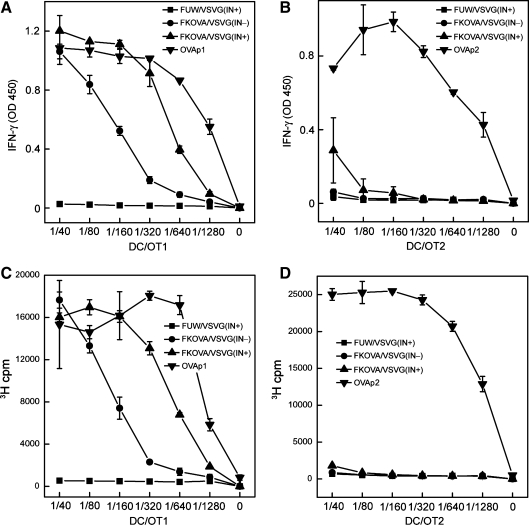
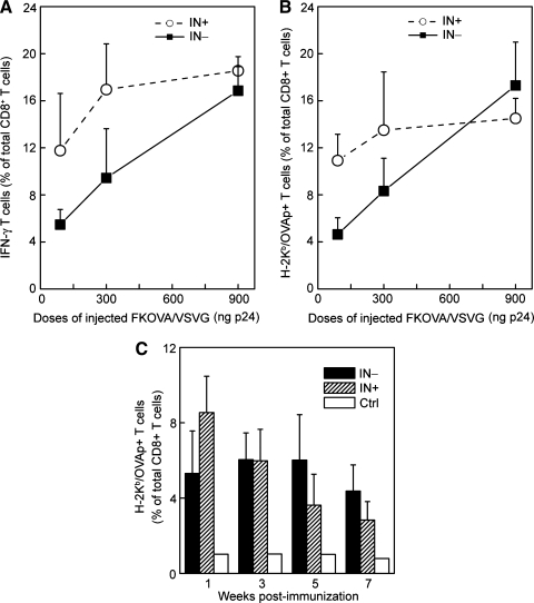
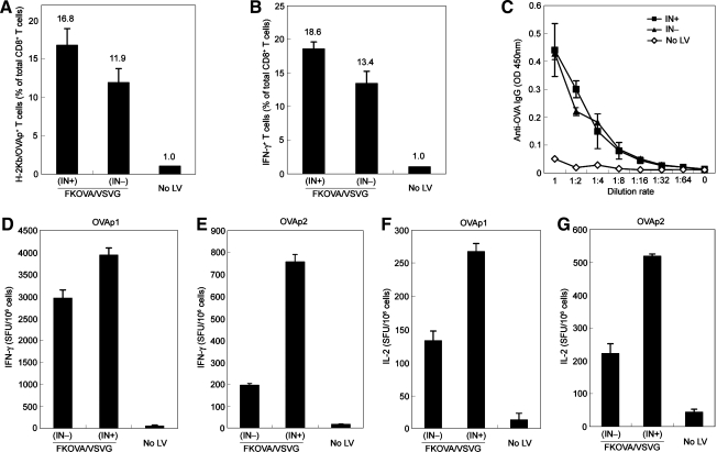
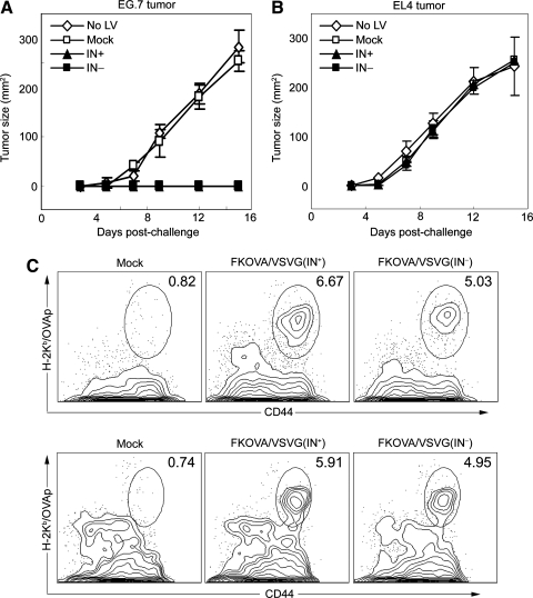
It has been demonstrated that nonintegrating lentiviral vectors (NILVs) are efficient in maintaining transgene expression in vitro and in vivo. Gene delivery by NILVs can significantly reduce nonspecific vector integration, which has been shown to cause malignant transformation in patients receiving gene therapy for X-linked severe combined immunodeficiency. Strong and sustained immune responses were observed after a single immunization with NILVs carrying viral antigens. However, there is no report to date that evaluates the efficacy of NILVs in inducing antigen-specific antitumor immunity. Using a well-characterized tumor model, we tested in vivo immunization with a self-inactivating lentiviral vector harboring a defective integrase. A high frequency of ovalbumin peptide (OVAp1)-specific CD8(+) T cells and a substantial antibody response were detected in naive mice immunized with an NILV encoding an OVA transgene. Furthermore, this immunization method completely protected the mice against the growth of E.G7 tumor cells expressing the OVA antigen. Thus, this study provides evidence that immunization using NILVs can be a safe and promising approach for exploring cancer immunotherapy.
Figures
Similar articles
-
Potent vaccine therapy with dendritic cells genetically modified by the gene-silencing-resistant retroviral vector GCDNsap.Mol Ther. 2006 Feb;13(2):301-9. doi: 10.1016/j.ymthe.2005.09.021. Epub 2005 Nov 28. Mol Ther. 2006. PMID: 16311073
-
Vaccines delivered by integration-deficient lentiviral vectors targeting dendritic cells induces strong antigen-specific immunity.Vaccine. 2010 Sep 24;28(41):6675-83. doi: 10.1016/j.vaccine.2010.08.012. Epub 2010 Aug 13. Vaccine. 2010. PMID: 20709004 Free PMC article.
-
Immunization with a lentiviral vector stimulates both CD4 and CD8 T cell responses to an ovalbumin transgene.Mol Ther. 2006 Feb;13(2):310-9. doi: 10.1016/j.ymthe.2005.08.025. Epub 2005 Nov 4. Mol Ther. 2006. PMID: 16275163
-
From pathogen to medicine: HIV-1-derived lentiviral vectors as vehicles for dendritic cell based cancer immunotherapy.J Gene Med. 2006 Jan;8(1):3-17. doi: 10.1002/jgm.846. J Gene Med. 2006. PMID: 16288497 Review.
-
The Old and the New: Prospects for Non-Integrating Lentiviral Vector Technology.Viruses. 2020 Sep 29;12(10):1103. doi: 10.3390/v12101103. Viruses. 2020. PMID: 33003492 Free PMC article. Review.
Cited by
-
Generation of a stable packaging cell line producing high-titer PPT-deleted integration-deficient lentiviral vectors.Mol Ther Methods Clin Dev. 2015 Jul 22;2:15025. doi: 10.1038/mtm.2015.25. eCollection 2015. Mol Ther Methods Clin Dev. 2015. PMID: 26229972 Free PMC article.
-
Nonintegrating Lentiviral Vector-Based Vaccine Efficiently Induces Functional and Persistent CD8+ T Cell Responses in Mice.J Biomed Biotechnol. 2010;2010:534501. doi: 10.1155/2010/534501. Epub 2010 May 19. J Biomed Biotechnol. 2010. PMID: 20508727 Free PMC article.
-
Pros and Cons of Antigen-Presenting Cell Targeted Tumor Vaccines.J Immunol Res. 2015;2015:785634. doi: 10.1155/2015/785634. Epub 2015 Oct 25. J Immunol Res. 2015. PMID: 26583156 Free PMC article. Review.
-
LV305, a dendritic cell-targeting integration-deficient ZVex(TM)-based lentiviral vector encoding NY-ESO-1, induces potent anti-tumor immune response.Mol Ther Oncolytics. 2016 Mar 30;3:16010. doi: 10.1038/mto.2016.10. eCollection 2016. Mol Ther Oncolytics. 2016. PMID: 27626061 Free PMC article.
-
Development of safer gene delivery systems to minimize the risk of insertional mutagenesis-related malignancies: a critical issue for the field of gene therapy.ISRN Oncol. 2012;2012:616310. doi: 10.5402/2012/616310. Epub 2012 Nov 22. ISRN Oncol. 2012. PMID: 23209944 Free PMC article.
References
-
- Apolonia L. Waddington S.N. Fernandes C. Ward N.J. Bouma G. Blundell M.P. Thrasher A.J. Collins M.K. Philpott N.J. Stable gene transfer to muscle using non-integrating lentiviral vectors. Mol. Ther. 2007;15:1947–1954. - PubMed
-
- Banchereau J. Palucka A.K. Dendritic cells as therapeutic vaccines against cancer. Nat. Rev. Immunol. 2005;5:296–306. - PubMed
-
- Banchereau J. Steinman R.M. Dendritic cells and the control of immunity. Nature. 1998;392:245–252. - PubMed
-
- Barnden M.J. Allison J. Heath W.R. Carbone F.R. Defective TCR expression in transgenic mice constructed using cDNA-based α- and β-chain genes under the control of heterologous regulatory elements. Immunol. Cell Biol. 1998;76:34–40. - PubMed
Publication types
MeSH terms
Substances
Grants and funding
LinkOut - more resources
Full Text Sources
Medical
Research Materials
