. 2009 Aug 7:10:367.
doi: 10.1186/1471-2164-10-367.

Complexity of the MSG gene family of Pneumocystis carinii

Affiliations

Complexity of the MSG gene family of Pneumocystis carinii

Scott P Keely et al. BMC Genomics. .

Abstract

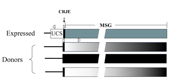
Background: The relationship between the parasitic fungus Pneumocystis carinii and its host, the laboratory rat, presumably involves features that allow the fungus to circumvent attacks by the immune system. It is hypothesized that the major surface glycoprotein (MSG) gene family endows Pneumocystis with the capacity to vary its surface. This gene family is comprised of approximately 80 genes, which each are approximately 3 kb long. Expression of the MSG gene family is regulated by a cis-dependent mechanism that involves a unique telomeric site in the genome called the expression site. Only the MSG gene adjacent to the expression site is represented by messenger RNA. Several P. carinii MSG genes have been sequenced, which showed that genes in the family can encode distinct isoforms of MSG. The vast majority of family members have not been characterized at the sequence level.

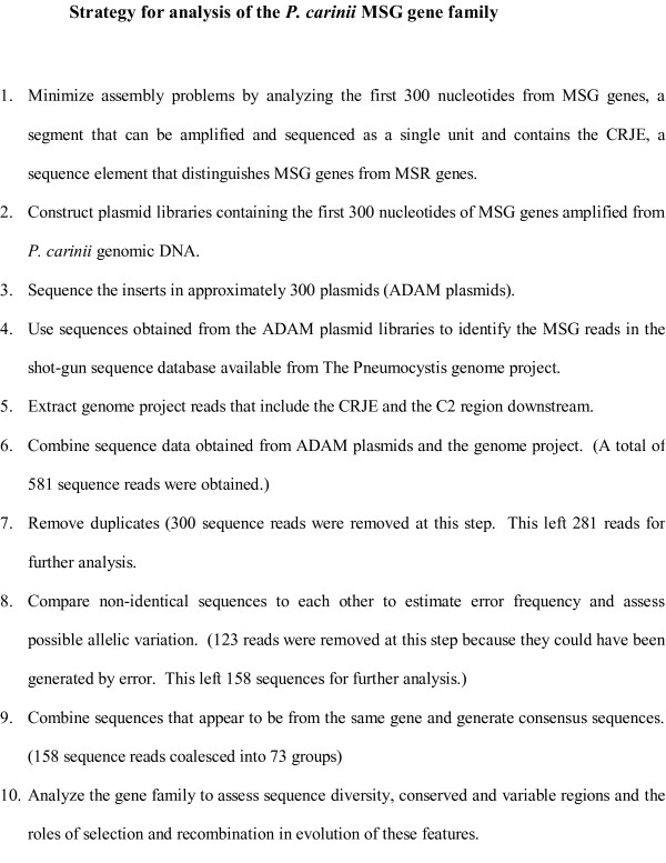
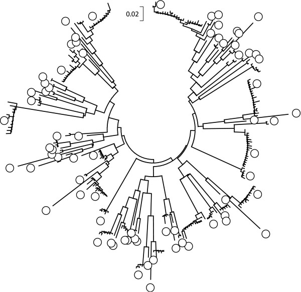
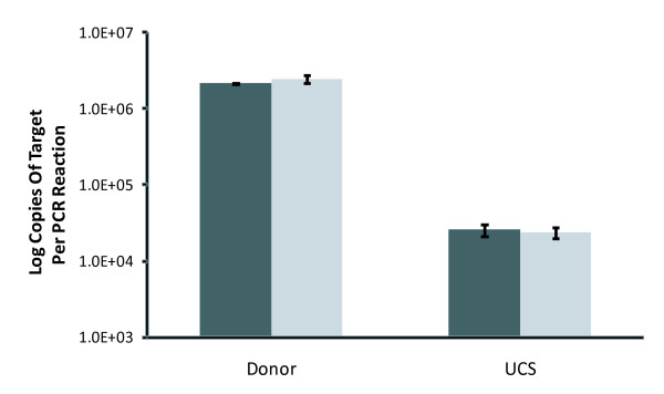
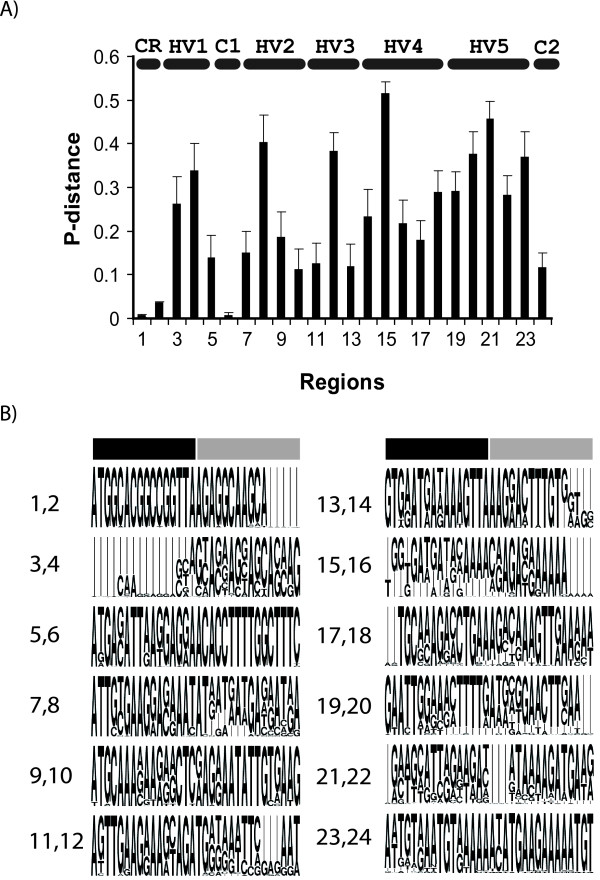
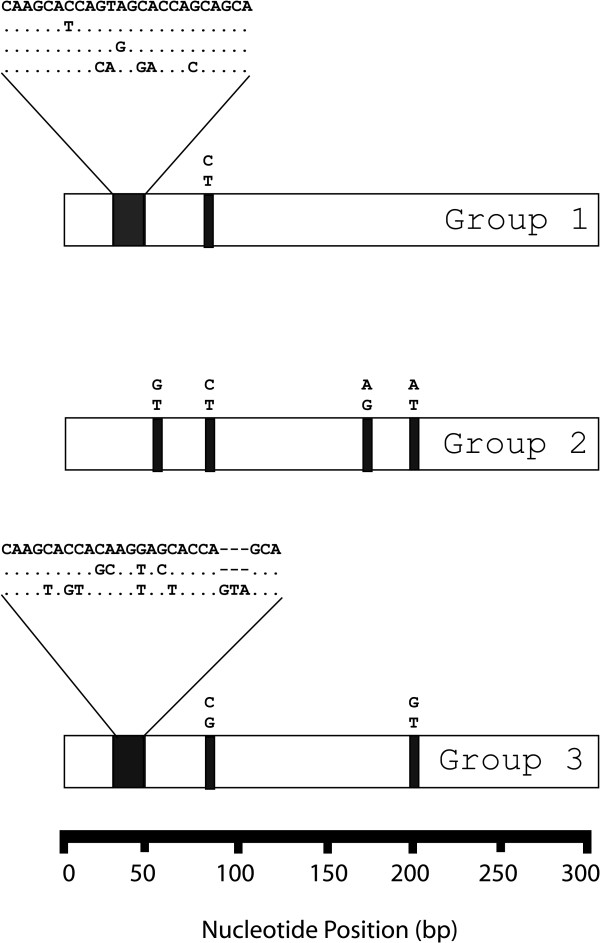
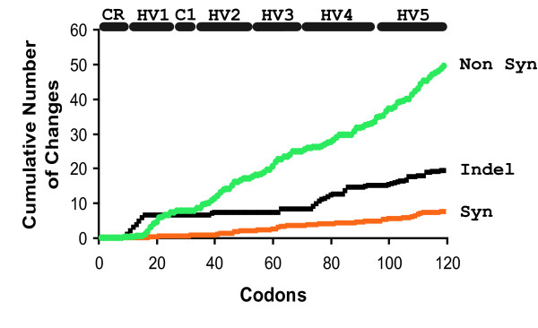
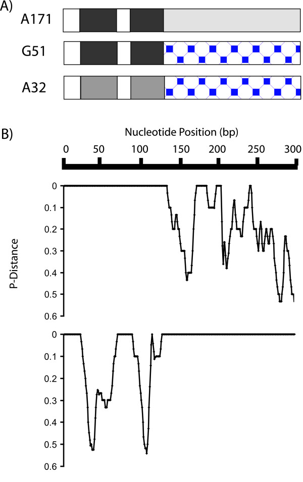
Results: The first 300 basepairs of MSG genes were subjected to analysis herein. Analysis of 581 MSG sequence reads from P. carinii genomic DNA yielded 281 different sequences. However, many of the sequence reads differed from others at only one site, a degree of variation consistent with that expected to be caused by error. Accounting for error reduced the number of truly distinct sequences observed to 158, roughly twice the number expected if the gene family contains 80 members. The size of the gene family was verified by PCR. The excess of distinct sequences appeared to be due to allelic variation. Discounting alleles, there were 73 different MSG genes observed. The 73 genes differed by 19% on average. Variable regions were rich in nucleotide differences that changed the encoded protein. The genes shared three regions in which at least 16 consecutive basepairs were invariant. There were numerous cases where two different genes were identical within a region that was variable among family members as a whole, suggesting recombination among family members.

Conclusion: A set of sequences that represents most if not all of the members of the P. carinii MSG gene family was obtained. The protein-changing nature of the variation among these sequences suggests that the family has been shaped by selection for protein variation, which is consistent with the hypothesis that the MSG gene family functions to enhance phenotypic variation among the members of a population of P. carinii.


Figures

Figure 1
Figure 1
Maps of expressed and donor MSG genes. The expressed MSG gene is adjacent to the UCS sequence. Donor MSG genes are not adjacent to the UCS sequence, which is unique in the genome. For illustrative purposes, just three of approximately 80 donor genes are depicted. There is a copy of the CRJE, which is a 24 basepair conserved sequence, at the beginning of MSG genes, including the one attached to the UCS. The discontinuities in the MSG genes are there to indicate that the genes are not drawn to size relative to the UCS and CRJE. The open horizontal arrows show the locations and orientations of PCR primers. The two arrows above the expressed gene represent the -145 and α-CRJE primers (Additional file 3, Table S2). The two arrows above the first donor gene represent the CRJE and C2 primers (Additional file 3, Table S2).
Figure 2
Figure 2
Strategy for analysis of the P. carinii MSG gene family.
Figure 3
Figure 3
Neighbor-Joining Tree of 281 MSG sequences. The relationships among the 281 non-identical MSG sequences were inferred using the Neighbor-Joining method [121] in MEGA4 software [117]. Gaps were ignored. Circle symbols mark the branches containing members of the 73 core MSG sequences (see text). The tree is drawn to scale in p-distance units. The bar at the top of the tree represents a p-distance of 0.02.
Figure 4
Figure 4
Quantification of the number of MSG genes by real-time PCR experiments. Donor genes were amplified using primers CRJE-RT and C1 (Additional file 3, Table S2). The UCS was amplified with UCS primer -145 and primer α-CRJE (Additional file 3, Table S2). At least three independent PCR reactions were performed with a given primer pair. The two bars of different shades show data obtained from P. carinii populations isolated from two different rats.
Figure 5
Figure 5
Conserved and variable regions in MSG genes. A) The 73 core MSG sequences were aligned. The DNA alignment was partitioned into regions containing 16 bp and average p-distances were calculated for each region using MEGA4 software [117]. The horizontal lines labeled HV1 etc, demarcate five hypervariable regions. The horizontal lines labeled CR, C1 and C2, demarcate constant regions CRJE, C1 and C2. B) A depiction of the majority and minority sequences observed. The height of a letter is proportionate to the frequency at which the base it represents was observed in the 73 aligned sequences. Thin vertical lines represent positions where INDELS occurred in the alignment. Each of the 12 blocks of sequence contains two of the twenty-four 16-base segments analyzed in panel A. The limits of each 16-base segment are indicated by the black and gray horizontal bars. The leftmost pairs of numbers correspond to the region-numbers in panel A. For example, the first block of 32 bases contains regions 1 and 2. Region 1 is covered by the black bar. Region 2 is covered by the gray bar.
Figure 6
Figure 6
Location and types of variation exhibited by closely-related sequence reads. Data derived from the top three groups of sequence reads (Table 1) are shown. An open bar represents a group of aligned sequences. The black boxes within an open bar indicate the locations where variable bases were observed among the reads in the group. The sequences observed are shown above each black box. Dots represent identity.
Figure 7
Figure 7
Positive selection in MSG genes. MSG genes were aligned and the frequency of non synonymous (abbreviated Non syn) and synonymous (Syn) substitutions as well as INDELS was scored for each codon. The horizontal lines labeled HV1 etc, demarcate five hypervariable regions. The horizontal lines labeled CR and C1 demarcate constant regions CRJE and C1.
Figure 8
Figure 8
Examples of gene structures suggestive of recombination between MSG genes. A171, G51 and A32 are three MSG sequences. A) Box diagram showing regions of identity among the three sequences. Regions that are identical for at least 16 basepairs are the same shade and pattern. B) Plots of identity (indicated by p-Distance = 0) and non-identity (indicated by p-Distances greater than zero) in pair-wise alignments of sequences G51 and A171 (upper) and G51 and A32 (lower). P-distances were calculated using a window size of 10 nucleotides and a step size of 1 nucleotide.

References

    1. Stringer JR, Edman JC, Cushion MT, Richards FF, Watanabe J. The fungal nature of Pneumocystis. J Med Vet Mycol. 1992;30:271–278. doi: 10.1080/02681219280000961. - DOI - PMC - PubMed
    1. Stringer SL, Stringer JR, Blase MA, Walzer PD, Cushion MT. Pneumocystis carinii: sequence from ribosomal RNA implies a close relationship with fungi. Exp Parasitol. 1989;68:450–461. doi: 10.1016/0014-4894(89)90130-6. - DOI - PubMed
    1. Redhead SA, Cushion MT, Frenkel JK, Stringer JR. Pneumocystis and Trypanosoma cruzi: Nomenclature and Typifications. J Eukaryot Microbiol. 2006;53:2–11. doi: 10.1111/j.1550-7408.2005.00072.x. - DOI - PubMed
    1. Aliouat-Denis CM, Chabe M, Demanche C, Aliouat eM, Viscogliosi E, Guillot J, et al. Pneumocystis species, co-evolution and pathogenic power. Infect Genet Evol. 2008;8:708–726. doi: 10.1016/j.meegid.2008.05.001. - DOI - PubMed
    1. Icenhour CR, Rebholz SL, Collins MS, Cushion MT. Widespread Occurrence of Pneumocystis carinii in Commercial Rat Colonies Detected Using Targeted PCR and Oral Swabs. J Clin Microbiol. 2001;39:3437–3441. doi: 10.1128/JCM.39.10.3437-3441.2001. - DOI - PMC - PubMed

Publication types

LinkOut - more resources