. 2009 Aug 7:7:48.
doi: 10.1186/1741-7007-7-48.

Induction of protein body formation in plant leaves by elastin-like polypeptide fusions

Affiliations

Induction of protein body formation in plant leaves by elastin-like polypeptide fusions

Andrew J Conley et al. BMC Biol. .

Abstract

Background: Elastin-like polypeptides are synthetic biopolymers composed of a repeating pentapeptide 'VPGXG' sequence that are valuable for the simple non-chromatographic purification of recombinant proteins. In addition, elastin-like polypeptide fusions have been shown to enhance the accumulation of a range of different recombinant proteins in plants, thus addressing the major limitation of plant-based expression systems, which is a low production yield. This study's main objectives were to determine the general utility of elastin-like polypeptide protein fusions in various intracellular compartments and to elucidate elastin-like polypeptide's mechanism of action for increasing recombinant protein accumulation in the endoplasmic reticulum of plants.

Results: The effect of elastin-like polypeptide fusions on the accumulation of green fluorescent protein targeted to the cytoplasm, chloroplasts, apoplast, and endoplasmic reticulum was evaluated. The endoplasmic reticulum was the only intracellular compartment in which an elastin-like polypeptide tag was shown to significantly enhance recombinant protein accumulation. Interestingly, endoplasmic reticulum-targeted elastin-like polypeptide fusions induced the formation of a novel type of protein body, which may be responsible for elastin-like polypeptide's positive effect on recombinant protein accumulation by excluding the heterologous protein from normal physiological turnover. Although expressed in the leaves of plants, these novel protein bodies appeared similar in size and morphology to the prolamin-based protein bodies naturally found in plant seeds. The elastin-like polypeptide-induced protein bodies were highly mobile organelles, exhibiting various dynamic patterns of movement throughout the cells, which were dependent on intact actin microfilaments and a functional actomyosin motility system.

Conclusion: An endoplasmic reticulum-targeted elastin-like polypeptide fusion approach provides an effective strategy for depositing large amounts of concentrated heterologous protein within the limited space of the cell via storage in stable protein bodies. Furthermore, encapsulation of recombinant proteins into physiologically inert organelles can function to insulate the protein from normal cellular mechanisms, thus limiting unnecessary stress to the host cell. Since elastin-like polypeptide is a mammalian-derived protein, this study demonstrates that plant seed-specific factors are not required for the formation of protein bodies in vegetative plant tissues, suggesting that the endoplasmic reticulum possesses an intrinsic ability to form protein body-like accretions in eukaryotic cells when overexpressing particular proteins.


Figures

Figure 1
Figure 1
Schematic representation of the genetic constructs used for Agrobacterium-mediated transient expression in Nicotiana benthamiana leaves. All transgene expression fragments were placed under the control of the cauliflower mosaic virus 35S promoter, a tCUP 5'-untranslated region and the nopaline synthase terminator. RuB-TP, transit peptide from the tobacco small subunit RuBisCo gene; Pr1b-SP, tobacco secretory signal peptide; N-glyco, N-glycosylation motif (GELVSNGTVT); BiP, tobacco immunoglobulin heavy chain binding protein; GFP, green fluorescent protein; YFP, yellow fluorescent protein; CFP, cyan fluorescent protein; ELP, elastin-like polypeptide tag (28×VPGVG); KDEL, endoplasmic reticulum retention signal.
Figure 2
Figure 2
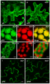
Subcellular localization of green fluorescent protein and green fluorescent protein-elastin-like polypeptide targeted to the cytoplasm, chloroplasts and apoplast. The localization constructs were agro-infiltrated into Nicotiana benthamiana leaves and visualized by confocal microscopy. The pG (A) and pGE (B) proteins were both visible as diffuse expression surrounding variously shaped and sized organelles throughout the cell. Green fluorescent protein (GFP) fluorescence was most concentrated in the cytoplasmic strands and the nucleus. In mesophyll cells expressing chloroplast-targeted GFP (pRG), the GFP fluorescence was localized to the chloroplasts (C), which was confirmed by also detecting the chlorophyll autofluorescence (D). (E) Merged image of (C) and (D) showing complete co-localization of pRG and the chloroplasts. In the presence of an elastin-like polypeptide (ELP) fusion tag, the chloroplast-targeted GFP (pRGE) appeared to accumulate in the cytoplasm (F) and was excluded from the chloroplasts (G). (H) Merged image of (F) and (G), demonstrating that an ELP fusion tag prevents the accumulation of GFP in the chloroplasts. For both pPG (I) and pPGE (J), the images were taken from a cross-section of the cells showing a secreted pattern of fluorescence consistent with apoplast localization. Bar, 10 μm (A, B, I, J); 5 μm (C to H).
Figure 3
Figure 3
Accumulation of green fluorescent protein in various subcellular compartments, in the presence or absence of an elastin-like polypeptide tag. The concentration of green fluorescent protein (GFP) was measured by quantitative fluorometric analysis from leaf sectors harvested from Nicotiana benthamiana plants 4 days post-agro-infiltration. Each column represents the mean value (n = 8), and the standard error of the mean is represented with error bars. Columns not connected with the same letter are significantly different (P < 0.05) from each other using Tamhane's T2 test. TSP, total soluble protein.
Figure 4
Figure 4
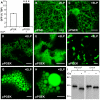
Hyperexpression of an endoplasmic reticulum-targeted elastin-like polypeptide fusion induces the formation of protein bodies in leaves. (A) Accumulation of endoplasmic reticulum- (ER-)targeted green fluorescent protein (GFP), with or without an elastin-like polypeptide (ELP) tag, when transiently co-expressed with the p19 suppressor of gene silencing in the leaves (n = 8) of Nicotiana benthamiana plants. ***, significant difference (P < 0.001). (B) Confocal image of the ER-targeted GFP control protein (pPGK) demonstrating the open polygonal network consistent with ER-localization. (C-H) In the presence of an ELP fusion tag, the ER-targeted GFP (pPGEK) was detected in brightly fluorescing spherical-shaped particles distributed throughout the cells of the leaf. (C) The novel PBs were closely associated with the ER tubules as small punctuate structures early on in the PB-formation process. (D) With time, the PB-like organelles continued to grow and appeared to be released from the ER into the cytoplasm, where they remained. (E-H) The PBs obtained various sizes and tended to cluster together within the cell, although the distribution pattern was quite variable. The majority of PBs had an observable diameter of between 0.5 and 1.0 μm, but larger PBs were seen at lower frequencies with some approaching diameters of 8.0 μm. (I) Deglycosylation of an ER-targeted GFP-ELP fusion engineered to contain an N-glycosylation motif (GELVSNGTVT). Total protein extracts (3 μg/lane) from agro-infiltrated plant tissue expressing pPNGEK were incubated for 24 h in the presence (+) or absence (-) of peptide N-glycosidase F (PNGaseF) or endoglycosidase H (EndoH) and then subjected to sodium dodecylsulphate-polyacrylamide gel electrophoresis and immunoblotted with an anti-GFP antibody. Bar, 10 μm (B-F); 5 μm (G, H).
Figure 5
Figure 5
Co-alignment of protein bodies with actin microfilaments. (A) Expression of the mouse talin (mTalin) actin-binding domain fused with YFP localizes to the actin cables of the cytoskeleton within leaf epidermal cells after agro-infiltration. (B) An endoplasmic reticulum-targeted cyan fluorescent protein-elastin-like polypeptide fusion (pPCEK) accumulated as protein bodies (PBs) within the cell's cytoplasm. (C-E) When co-expressed, the induced novel PBs co-aligned with the actin microfilaments. Bar, 10 μm.
Figure 6
Figure 6
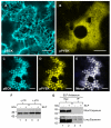
Endoplasmic reticulum luminal binding protein is localized to the novel protein bodies, but does not specifically interact with elastin-like polypeptide. (A) The tobacco endoplasmic reticulum- (ER-)resident chaperone binding protein (BiP) fused with cyan fluorescent protein (CFP) (pBCK) is appropriately localized to the ER. (B) An ER-targeted yellow fluorescent protein-elastin-like polypeptide (YFP-ELP) fusion (pPYEK) accumulated in the induced protein bodies (PBs) located in the cytoplasm. (C-E) When co-expressed, pBCK co-localized with the PBs induced by pPYEK expression, suggesting that the novel PBs originate from the ER. (F) Western blot analysis comparing BiP accumulation in leaves transiently expressing ER-targeted GFP in the absence (pPGK) or presence (pPGEK) of an ELP tag, with or without co-agro-infiltration of the p19 suppressor of gene silencing. Total protein extracts (30 μg/lane) were resolved by sodium dodecylsulphate-polyacrylamide gel electrophoresis (SDS-PAGE), followed by immunoblotting using an anti-BiP antibody. (G) Leaf extracts expressing pPGK or pPGEK were immunoprecipitated with anti-ELP antiserum. The supernatants (Sup) and immunoprecipitates (IP) were separated by SDS-PAGE and probed with anti-BiP antibody. Bar, 10 μm (A, B); 5 μm (C-E).
Figure 7
Figure 7
Subcellular localization of the endoplasmic reticulum-targeted green fluorescent protein-elastin-like polypeptide fusion protein (pPGEK) in Nicotiana benthamiana leaves. (A) Electron microscopy confirmed the location of numerous newly-formed endoplasmic reticulum- (ER-)derived protein bodies (PBs) (examples indicated by asterisks) in the cytoplasm of the leaf cells. (B-D) Progressively higher magnifications of the PBs seen in (A). (B) The novel PBs occupied the cytosolic space between the tonoplast (indicated by an arrowhead) and the plasma membrane (indicated by an arrow). (C) The PBs were clearly surrounded by a membrane that appears to no longer be contiguous with the ER. (D) The PB membrane was decorated with ribosomes (indicated with arrows), strongly suggesting that the PBs were originally derived from the rough ER. (E, F) Immunogold localization confirmed the presence of green fluorescent protein-elastin-like polypeptide (GFP-ELP) inside the novel cytoplasmic PBs in ultrathin sections of N. benthamiana leaves using anti-ELP (E) and anti-GFP (F) antibodies detected with goat anti-rabbit or anti-mouse IgG conjugated to 15 nm gold particles. No significant immunolabeling was observed in other subcellular compartments or wild-type plants. The different subcellular compartments were labeled: Cp, chloroplast; CW, cell wall; Mt, mitochondria; LB, lipid body; LV, lytic vacuole; *, induced protein body. Bar, 2 μm (A); 500 nm (B, C, E, F); 100 nm (D).

Similar articles

Cited by

References

    1. Stoger E, Ma JK, Fischer R, Christou P. Sowing the seeds of success: Pharmaceutical proteins from plants. Curr Opin Biotechnol. 2005;16:167–173. - PubMed
    1. Galili G. ER-derived compartments are formed by highly regulated processes and have special functions in plants. Plant Physiol. 2004;136:3411–3413. - PMC - PubMed
    1. Arcalis E, Marcel S, Altmann F, Kolarich D, Drakakaki G, Fischer R, Christou P, Stoger E. Unexpected deposition patterns of recombinant proteins in post-endoplasmic reticulum compartments of wheat endosperm. Plant Physiol. 2004;136:3457–3466. - PMC - PubMed
    1. Herman EM, Larkins BA. Protein storage bodies and vacuoles. Plant Cell. 1999;11:601–614. - PMC - PubMed
    1. Geli MI, Torrent M, Ludevid D. Two structural domains mediate two sequential events in gamma-zein targeting: Protein endoplasmic reticulum retention and protein body formation. Plant Cell. 1994;6:1911–1922. - PMC - PubMed

Publication types

MeSH terms

LinkOut - more resources