

Microdeletion of 6q16.1 encompassing EPHA7 in a child with mild neurological abnormalities and dysmorphic features: case report
- PMID: 19664229
- PMCID: PMC2731778
- DOI: 10.1186/1755-8166-2-17
Microdeletion of 6q16.1 encompassing EPHA7 in a child with mild neurological abnormalities and dysmorphic features: case report
Abstract
Background: Of the fewer than 100 cases reported within the literature of constitutional deletions involving the long arm of chromosome 6, only five have been characterized using high-resolution microarray analysis. Reported 6q deletion patients show a high incidence of mental retardation, ear anomalies, hypotonia, and postnatal growth retardation.
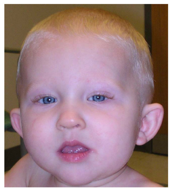
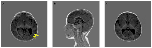
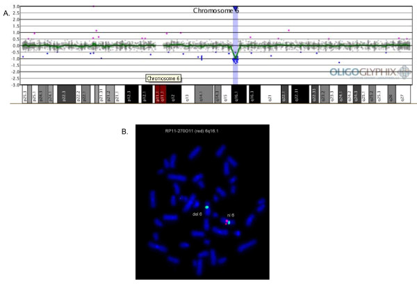
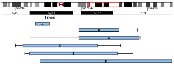
Results: We report a 16-month-old male presenting with developmental delay and dysmorphic features who was found by array-based comparative genomic hybridization (aCGH) to have a ~2.16 Mb de novo deletion within chromosome band 6q16.1 that encompasses only two genes. Expression studies of the mouse homologue of one of the genes, the ephrin receptor 7 gene (EPHA7), have shown the gene functions during murine embryogenesis to form cortical domains, determine brain size and shape, and play a role in development of the central nervous system (CNS).
Discussion: Our results suggest that deletion of EPHA7 plays a role in the neurologic and dysmorphic features, including developmental delay, hypotonia, and ear malformations, observed in some 6q deletion patients.
Figures
Similar articles
-
EPHA7 haploinsufficiency is associated with a neurodevelopmental disorder.Clin Genet. 2021 Oct;100(4):396-404. doi: 10.1111/cge.14017. Epub 2021 Jul 1. Clin Genet. 2021. PMID: 34176129
-
Whole-genome array-CGH identifies novel contiguous gene deletions and duplications associated with developmental delay, mental retardation, and dysmorphic features.Am J Med Genet A. 2007 Jul 1;143A(13):1431-41. doi: 10.1002/ajmg.a.31773. Am J Med Genet A. 2007. PMID: 17568414
-
Severe mental retardation, seizures, and hypotonia due to deletions of MEF2C.Am J Med Genet B Neuropsychiatr Genet. 2010 Jul;153B(5):1042-51. doi: 10.1002/ajmg.b.31071. Am J Med Genet B Neuropsychiatr Genet. 2010. PMID: 20333642
-
Interstitial 6q microdeletion syndrome and epilepsy: a new patient and review of the literature.Am J Med Genet A. 2013 Aug;161A(8):2009-15. doi: 10.1002/ajmg.a.35993. Epub 2013 Jun 21. Am J Med Genet A. 2013. PMID: 23794236 Review.
-
The discovery of microdeletion syndromes in the post-genomic era: review of the methodology and characterization of a new 1q41q42 microdeletion syndrome.Genet Med. 2007 Sep;9(9):607-16. doi: 10.1097/gim.0b013e3181484b49. Genet Med. 2007. PMID: 17873649 Review.
Cited by
-
Homozygous deletion of Tenascin-R in a patient with intellectual disability.J Med Genet. 2012 Jul;49(7):451-4. doi: 10.1136/jmedgenet-2012-100831. Epub 2012 Jun 22. J Med Genet. 2012. PMID: 22730557 Free PMC article.
-
Molecular and clinical delineation of the 17q22 microdeletion phenotype.Eur J Hum Genet. 2013 Oct;21(10):1085-92. doi: 10.1038/ejhg.2012.306. Epub 2013 Jan 30. Eur J Hum Genet. 2013. PMID: 23361222 Free PMC article.
-
Recurrent HERV-H-mediated 3q13.2-q13.31 deletions cause a syndrome of hypotonia and motor, language, and cognitive delays.Hum Mutat. 2013 Oct;34(10):1415-23. doi: 10.1002/humu.22384. Epub 2013 Aug 13. Hum Mutat. 2013. PMID: 23878096 Free PMC article.
-
Refinement of the Region for Split Hand/Foot Malformation 5 on 2q31.1.Mol Syndromol. 2010;1(5):262-271. doi: 10.1159/000328405. Epub 2011 May 18. Mol Syndromol. 2010. PMID: 22140379 Free PMC article.
-
Haploinsufficiency of SOX5 at 12p12.1 is associated with developmental delays with prominent language delay, behavior problems, and mild dysmorphic features.Hum Mutat. 2012 Apr;33(4):728-40. doi: 10.1002/humu.22037. Hum Mutat. 2012. PMID: 22290657 Free PMC article.
References
-
- Bonaglia MC, Ciccone R, Gimelli G, Gimelli S, Marelli S, Verheij J, Giorda R, Grasso R, Borgatti R, Pagone F, et al. Detailed phenotype-genotype study in five patients with chromosome 6q16 deletion: narrowing the critical region for Prader-Willi-like phenotype. Eur J Hum Genet. 2008;16:1443–1449. doi: 10.1038/ejhg.2008.119. - DOI - PubMed
LinkOut - more resources
Full Text Sources
Other Literature Sources
Miscellaneous
