. 2009 Aug 11:6:73.
doi: 10.1186/1742-4690-6-73.

Involvement of TORC2, a CREB co-activator, in the in vivo-specific transcriptional control of HTLV-1

Affiliations

Involvement of TORC2, a CREB co-activator, in the in vivo-specific transcriptional control of HTLV-1

Shiwen Jiang et al. Retrovirology. .

Abstract

Background: Human T-cell leukemia virus type 1 (HTLV-1) causes adult T -cell leukemia (ATL) but the expression of HTLV-1 is strongly suppressed in the peripheral blood of infected people. However, such suppression, which may explain the long latency in the development of ATL, is readily reversible, and viral expression resumes quickly with ex vivo culture of infected T -cells. To investigate the mechanism of in vivo -specific transcriptional suppression, we established a mouse model in which mice were intraperitoneally administered syngeneic EL4 T -lymphoma cells transduced with a recombinant retrovirus expressing a GFP-Tax fusion protein, Gax, under the control of the HTLV-1 enhancer (EL4-Gax).

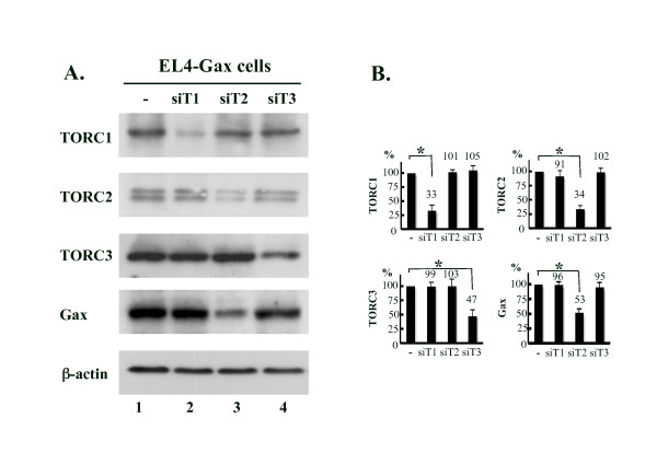
Results: Gax gene transcription was silenced in vivo but quickly up-regulated in ex vivo culture. Analysis of integrated Gax reporter gene demonstrated that neither CpG methylation of the promoter DNA nor histone modification was associated with the reversible suppression. ChIP-analysis of LTR under suppression revealed reduced promoter binding of TFIIB and Pol-II, but no change in the binding of CREB or CBP/p300 to the viral enhancer sequence. However, the expression of TORC2, a co-activator of CREB, decreased substantially in the EL4-Gax cells in vivo, and this returned to normal levels in ex vivo culture. The reduced expression of TORC2 was associated with translocation from the nucleus to the cytoplasm. A knock-down experiment with siRNA confirmed that TORC2 was the major functional protein of the three TORC-family proteins (TORC1, 2, 3) in EL4-Gax cells.

Conclusion: These results suggest that the TORC2 may play an important role in the in vivo -specific transcriptional control of HTLV-1. This study provides a new model for the reversible mechanism that suppresses HTLV-1 expression in vivo without the DNA methylation or hypoacetylated histones that is observed in the primary cells of most HTLV-1 -infected carriers and a substantial number of ATL cases.


Figures

Figure 1
Figure 1
A mouse model system with EL4-Gax cells. A. The structural organization of the R3Gaxbsr genome in EL4-Gax cells [29]. The EGFP coding sequence was fused with tax cDNA at the 5'-end, resulting in Gax. The Gax gene was linked with a drug resistance gene, bsr, by an internal ribosome entry site (IRES). The U3 region in the MLV LTR was replaced with that in the HTLV-1 LTR. 1 × 10⁶ of EL4-Gax cells cultured in vitro (in vitro, a) were injected into peritoneal cavity of a syngenic C57BL/6J mouse. 3 weeks after challenge, cells were collected from ascitic fluids (in vivo, b) and transferred to the in vitro culture condition for 48 hours (ex vivo, c). B. Left, the expression of Gax protein in living cells was monitored as the intensity of GFP fluorescence by fluorescent-activated cell sorter (FACS); Right, statistical analysis of the GFP mean fluorescent intensity (GFP mfi), after deducting EL4 cells background level. EL4, parent cell line. C. Left, cell lysates were subjected to Western blot analysis with anti-Tax serum, PARP, an indicator of apoptosis or necrosis exhibiting the signature 89 kDa or 50 kDa fragment respectively to see if the in vivo cells were healthy or not, or anti-β-actin antibody as loading control; Right, quantification of Gax and normalized to β-actin with a densitometry software program (NIH-image). D. Left, RT-PCR analysis of several viral and cellular mRNAs.; Right, Real-time PCR analysis of Gax mRNA expression and normalized to 18S ribosomal RNA in EL4-Gax cells with SYBR Green. Error bars indicate SEMs. Data were obtained from four independent experiments analyzing one mouse per experiment, and statistical analysis of the data was performed between the in vivo or ex vivo against the in vitro. *; p < 0.01. **; p < 0.05.
Figure 2
Figure 2
CpG methylation of the enhancer/promoter region of provirus DNA in EL4-Gax cells. Top: locations of CpG sites (#1–11) in the HTLV-1 U3 region studied in this experiment. The sense primer is complementary to the mouse genomic sequence flanking the 5'-LTR of provirus at the integration site, and the anti-sense primer is complementary to the junction sequence between the HTLV-1 and MLV U3 regions. The three 21 -bp enhancer sequences are indicated as boxes. Bottom: results of bisulfite genomic sequencing analysis of four independent experiments. Methylated and unmethylated CpG sites are expressed as filled and open rectangles, respectively. Amplified PCR products were subcloned into pGEM-T vector, and the nucleotide sequences of at least 13 clones were determined. GFP mfi: the GFP mean fluorescent intensity of EL4-Gax cells used for bisulfite genomic sequencing analysis.
Figure 3
Figure 3
ChIP analysis of the enhancer/promoter region in Gax provirus. A. Schematic representation of the 5'-LTR in the R3Gaxbsr reporter gene. The three 21 -bp enhancer sequences (boxes), the TATA sequence, and the transcription start site (+1) are shown. Primers for PCR are indicated by arrows. The 5'- and 3'-ends of amplified DNA are denoted as the nucleotide positions relative to the transcription start site. Primers #1 and #2 amplify the enhancer region, and primers #3 and #4 amplify the promoter region of Gax-5'-LTR. B. Binding of CREB, phosphor-CREB and CBP to the Gax enhancer region was constant in EL4-Gax cells in vitro (a) and in vivo (b)(left), but the enhancer binding of Gax was reduced when EL4-Gax cells were grown in vivo (right). C. Expression of CREB protein in EL4-Gax cells. Anti-CREB1, anti-phosphor-CREB antibodies were used to detect proteins in EL4-Gax cells grown in vitro, in vivo, and ex vivo. Equivalent protein loading was confirmed by stripping and re-probing the blot with an anti-β-actin antibody. D. Binding of acetylated histone 3 at Lys-9, 14 (H3), acetylated histone 4 at Lys- 5, 8, 12, 16 (H4) and trimethylated histone 3 at Lys-4 (H3K4tri) to the Gax enhancer region was not changed in EL4-Gax cells either in vitro (a) or in vivo (b), but the promoter binding of the basic transcription factor TFIIB and of RNA polymerase II (Pol-II) was reduced when EL4-Gax cells were grown in vivo. Factors binding to promoters of EF-1a and β-globin are presented as positive and negative controls, respectively.
Figure 4
Figure 4
Expression of TORC proteins in EL4-Gax cells. Anti-TORC1 (A), anti-TORC2 (B), and anti-TORC3 (C) antibodies were used to detect each protein in EL4-Gax cells grown in vitro, in vivo, and ex vivo. Equivalent protein loading was confirmed by stripping and re-probing the blot with an anti-β-actin antibody. Apparent molecular weights of marker protein are indicated. D. Phosphorylation of TORC2. Protein from EL4-Gax cells was incubated with or without rAPid Alkaline Phosphatase (see methods in detail). E. ChIP analysis of TORCs in EL4-Gax cells in vitro (a) and in vivo (b). Little or no binding of TORC1 and TORC3 to the Gax enhancer region was observed in vitro (a) or in vivo (b), but the binding of TORC2 to the Gax enhancer region was high in vitro (a) and reduced when EL4-Gax cells were grown in vivo (b).
Figure 5
Figure 5
Effect of the knock-down of TORC genes on the expression of Gax in EL4-Gax cells. A. Expression of TORC proteins and Gax in EL4-Gax cells transduced with retrovirus (for TORC1 and 3) or lentivirus (for TORC2) vectors encoding siRNA against each TORC gene. Expression of the siRNA resulted in the reduction of respective TORC proteins, but only siRNA to the TORC2 gene suppressed Gax gene expression. Proteins were detected with antibodies to the respective TORC proteins, Tax, or β-actin by the ECL or ECL plus system. B. Densitometric analysis data of TORC proteins and Gax protein normalized to β-actin are presented as mean ± SEM of three independent experiments. T1, TORC1; T2, TORC2; T3, TORC3.
Figure 6
Figure 6
Expression and subcellular localization of TORC2 in EL4-Gax cells. A. Immunofluorescent staining of TORC2 protein (red) in EL4-Gax cells grown in vitro, in vivo, or ex vivo. Cells were counterstained with DAPI (blue) to localize the nucleus and examined by confocal microscopy. The right panel shows a magnified image of a single cell indicated by an arrow in the adjacent panel. B. Statistical analysis of subcellular localization of TORC2 expression. The amount of protein expression was determined as the number of fluorescent pixels in the total and nucleus for each growth condition. Data were obtained by counting pixels in 108, 137, and 106 cells for in vitro, in vivo, and ex vivo conditions, respectively. Error bars indicate SEMs. *; p < 0.001. C. Expression of AMPKα protein in EL4-Gax cells. Anti-AMPKα and anti-phosphor-AMPKα (Thr172) antibodies were used to detect protein in EL4-Gax cells grown in vitro, in vivo, and ex vivo. Equivalent protein loading was confirmed by stripping and re-probing the blot with an anti-β-actin antibody.

References

    1. Yoshida M, Miyoshi I, Hinuma Y. Isolation and characterization of retrovirus from cell lines of human adult T-cell leukemia and its implication in the disease. Proc Natl Acad Sci U S A . 1982;79:2031–2035. doi: 10.1073/pnas.79.6.2031. - DOI - PMC - PubMed
    1. Poiesz BJ, Ruscetti FW, Gazdar AF, Bunn PA, Minna JD, Gallo RC. Detection and isolation of type C retrovirus particles from fresh and cultured lymphocytes of a patient with cutaneous T-cell lymphoma. Proc Natl Acad Sci USA. 1980;77:7415–7419. doi: 10.1073/pnas.77.12.7415. - DOI - PMC - PubMed
    1. Osame M, Usuku K, Izumo S, Ijichi N, Amitani H, Igata A, Matsumoto M, Tara M. HTLV-I associated myelopathy, a new clinical entity. Lancet. 1986;1:1031–1032. doi: 10.1016/S0140-6736(86)91298-5. - DOI - PubMed
    1. Gessain A, Barin F, Vernant JC, Gout O, Maurs L, Calender A, de The G. Antibodies to human T-lymphotropic virus type-I in patients with tropical spastic paraparesis. Lancet. 1985;2:407–410. doi: 10.1016/S0140-6736(85)92734-5. - DOI - PubMed
    1. Seiki M, Hattori S, Hirayama Y, Yoshida M. Human adult T-cell leukemia virus: complete nucleotide sequence of the provirus genome integrated in leukemia cell DNA. Proc Natl Acad Sci USA. 1983;80:3618–3622. doi: 10.1073/pnas.80.12.3618. - DOI - PMC - PubMed

Publication types

LinkOut - more resources