

Update on amyloid imaging: from healthy aging to Alzheimer's disease
- PMID: 19664363
- PMCID: PMC2825106
- DOI: 10.1007/s11910-009-0051-4
Update on amyloid imaging: from healthy aging to Alzheimer's disease
Abstract
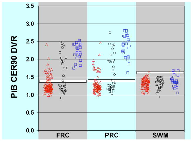
We report on the current state of in vivo amyloid imaging. Although this technique is less than a decade old, a wealth of information is emerging as the initial clinical studies are being reported. Imaging of patients with Alzheimer's disease has allowed quantitative exploration of the natural history of amyloid deposition and its relationship to neurodegeneration. Amyloid imaging also shows significant promise in the differential diagnosis of mild cognitive impairment or atypical dementias. However, amyloid detection may be of greatest utility in healthy elderly persons; consistent with prior autopsy studies, a significant proportion of asymptomatic individuals display increased levels of amyloid by in vivo imaging. Understanding the relationship between this pathology and future cognitive status has significant implications for the application of disease-modifying medications in the "preclinical" phase of disease. Given the considerable clinical experience compared with other tracers, this review focuses on the literature involving Pittsburgh compound B positron emission tomography.
Figures

References
-
- Small GW, Kepe V, Ercoli LM, et al. PET of brain amyloid and tau in mild cognitive impairment. N Engl J Med. 2006;355:2652–63. - PubMed
-
- Verhoeff NP, Wilson AA, Takeshita S, et al. In-vivo imaging of Alzheimer disease beta-amyloid with [11C]SB-13 PET. Am J Geriatr Psychiatry. 2004;12:584–95. - PubMed
-
- Klunk WE, Engler H, Nordberg A, et al. Imaging brain amyloid in Alzheimer's disease with Pittsburgh Compound-B. Ann Neurol. 2004;55:306–19. - PubMed
Publication types
MeSH terms
Substances
Grants and funding
LinkOut - more resources
Full Text Sources
Other Literature Sources
Medical