. 2009 Dec;162(1-2):133-41.
doi: 10.1016/j.jviromet.2009.07.028. Epub 2009 Aug 5.

Heterologous expression of full-length capsid protein of porcine circovirus 2 in Escherichia coli and its potential use for detection of antibodies

Affiliations

Heterologous expression of full-length capsid protein of porcine circovirus 2 in Escherichia coli and its potential use for detection of antibodies

Zuzana Marcekova et al. J Virol Methods. 2009 Dec.

Abstract

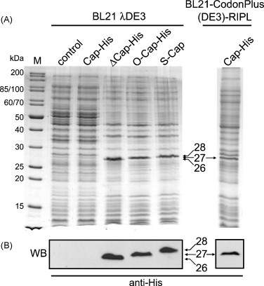
A capsid protein of porcine circovirus 2 (PCV 2) serves as a diagnostic antigen for the detection of PCV 2-associated disease known as a postweaning multisystemic wasting syndrome (PMWS). In this report, a bacterial expression system was developed for the expression and purification of the full-length PCV 2 capsid (Cap) protein from a codon-optimized cap gene. Replacement of rare arginine codons located at the 5' end of the cap reading frame with codons optimal for E. coli was found to overcome the poor expression of the viral protein in the prokaryotic system. The Cap protein was purified to greater than 95% homogeneity by using a single cation-exchange chromatography at a yield of 10 mg per litre of bacterial culture. Despite the failure of the E. coli-expressed Cap protein to self-assemble into virus-like particles (VLPs), the immunization of mice with recombinant Cap yielded antibodies with the same specificity as those raised against native PCV 2 virions. In addition, the antigenic properties of the purified Cap protein were employed in a subunit-based indirect ELISA to monitor the levels of PCV 2 specific antibodies in piglets originating from a herd which was experiencing PCV 2 infection. These results pave the way for a straightforward large-scale production of the recombinant PCV 2 capsid protein and its use as a diagnostic antigen or a PCV 2 subunit vaccine.


Figures

Fig. 1
Fig. 1
A scheme of constructs used for PCV 2 capsid (Cap) protein expression. (A) Primary amino acid sequence of the Cap protein. The nuclear localization signal domain is in the grey box, the N-terminal arginine residues are highlighted in bold italic. (B) Schematic representation of the Cap protein variants used in this study. The designation of the protein and its expression vector is indicated. (C) Codon optimization of the 5′ end of the cap gene. A comparison of the original cap gene nucleotide sequence with the sequence of codon-optimized (O-cap-His) and the fully synthetic gene (S-cap). The modified codons are underlined. The nucleotide sequence in the box represents the deleted codons (Δ) in ΔCap-His protein. The MscI restriction site in the original cap gene is indicated.
Fig. 2
Fig. 2
The expression of various Cap proteins in E. coli. (A) SDS-PAGE analysis of cellular proteins obtained from IPTG-induced E. coli cells. M, molecular weight markers (in kDa); control, control cells (no IPTG); Cap-His, expression of the Cap-His protein; ΔCap-His, expression of the ΔCap-His protein; O-Cap-His, expression of the O-Cap-His protein; S-Cap, expression of the S-Cap protein. The expression of the Cap-His protein in E. coli BL21-CodonPlus(DE3)-RIPL is indicated on the right. (B) Western blot analysis of total cellular proteins obtained from IPTG-induced E. coli cells by using anti-His antibody. The blot corresponds to lanes 2–6 from (A). The arrows represent the location of the ΔCap-His (26 kDa), O-Cap-His (27 kDa), S-Cap (28 kDa), and Cap-His proteins, respectively.
Fig. 3
Fig. 3
Purification of recombinant S-Cap protein. (A) UnoSphere-S cation-exchange elution profile of cytosolic extract of E. coli BL21 λ(DE3) expressing the S-Cap protein. The cytosolic extract was applied onto the column and the bound proteins were eluted with a continuous gradient of NaCl (dotted line). (B) SDS-PAGE analysis of fractions collected from the UnoSphere-S cation-exchange chromatography. M, molecular weight markers (in kDa). Protein profiles of individual elution fractions were indicated by a fraction number. (C) Western blot analysis of fractions collected from the UnoSphere-S cation-exchange chromatography by using anti-His antibody. The blot corresponds to lanes 2–8 from (B).
Fig. 4
Fig. 4
Virus-like particles (VLPs) formation in the cytosol of bacterial cells. Electron micrographs of ultrathin sections of E. coli BL21 λ(DE3) expressing a fusion protein of capsid and nucleocapsid protein (CANC) obtained from the Gag polyprotein of Mason-Pfizer monkey virus (Ulbrich et al., 2006) (A and B), and E. coli BL21 λ(DE3) expressing the Cap protein (C and D). (A) Non-induced cells, (B) IPTG-induced cells expressing CANC filled with spherical VLPs, (C) non-induced cells and (D) IPTG-induced cells expressing the Cap protein. No VLPs are visible in the cytosol. Note the presence of outer membrane vesicles at the exterior of the cells (arrowheads). Bar, 500 nm.
Fig. 5
Fig. 5
Specificity and sensitivity of the indirect Cap-ELISA. Different pig sera were diluted to 1:100 with PBS and their reactivity with the recombinant S-Cap and ΔCap-His proteins (10 μg/ml) was analyzed by a colorimetric reaction at the optical density at 450 nm (OD450nm). OD450nm of 0.2 represents a cut-off value for seropositivity of the samples. TGE-specific and PED-specific pig sera were obtained from SPF pigs which were experimentally infected with the coronavirus causing transmissible gastroenteritis (TGE) and porcine epidemic diarrhoea (PED), respectively. Data represent the means ± S.D. for the three independent experiments.
Fig. 6
Fig. 6
Time course analysis of levels of PCV 2-specific antibodies (A) and DNA copy numbers of PCV 2 per 1 ml of serum (B) in piglets in a herd with PCV2 infection during their first 20 weeks of age. (A) Time course analysis of the PCV 2-specific antibody levels as detected by the indirect Cap-ELISA. The final values were expressed as a signal-to-positive (S/P) ratio. S/P value was determined according to the following formula: (OD450nm of sample − OD450nm of negative control)/(OD450nm of positive control − OD450nm of negative control). S/P value of 0.3 represents the cut-off signal for seropositivity of the samples. (B) Time course analysis of the PCV2 DNA copy numbers per 1 ml of serum as detected by the quantitative PCR. Total genomic DNA was extracted from 200 μl of each serum sample and quantified by the PCR amplification of 100 bp DNA segment within the nucleotide sequence of the PCV 2 cap gene using a TaqMan format. Results of statistical analysis using one-way analysis of variance (ANOVA) between serum samples obtained at given time intervals from 22 piglets are represented by the box plots. The median value for each dataset is indicated by the black center line. The vertical height of each box indicates the 25–75% data range. The upper and lower bars denote the largest and smallest data values. The marker (○) denotes the extreme value of the individual serum sample that is >1.5 times the inter-quartile range from the upper and lower quartile. The marker (●) denotes the extreme value of the individual serum sample that is >3 times the inter-quartile range from the upper quartile. Data represent the means ± S.D. for the three independent experiments.

Similar articles

Cited by

References

    1. Allan G.M., Ellis J.A. Porcine circoviruses: a review. J. Vet. Diagn. Invest. 2000;12:3–14. - PubMed
    1. Allan G.M., McNeilly F., Kennedy S., Daft B., Clarke E.G., Ellis J.A., Haines D.M., Meehan B.M., Adair B.M. Isolation of porcine circovirus-like viruses from pigs with a wasting disease in the United States of America and Europe. J. Vet. Diagn. Invest. 1998;10:3–10. - PubMed
    1. Blanchard P., Mahe D., Cariolet R., Truong C., Le Dimna M., Arnauld C., Rose N., Eveno E., Albina E., Madec F., Jestin A. An ORF2 protein-based ELISA for porcine circovirus type 2 antibodies in post-weaning multisystemic wasting syndrome. Vet. Microbiol. 2003;94:183–194. - PMC - PubMed
    1. Brunborg I.M., Moldal T., Jonassen C.M. Quantitation of porcine circovirus type 2 isolated from serum/plasma and tissue samples of healthy pigs and pigs with postweaning multisystemic wasting syndrome using a TaqMan-based real-time PCR. J. Virol. Methods. 2004;122:171–178. - PubMed
    1. Burgess-Brown N.A., Sharma S., Sobott F., Loenarz C., Oppermann U., Gileadi O. Codon optimization can improve expression of human genes in Escherichia coli: a multi-gene study. Protein Expr. Purif. 2008;59:94–102. - PubMed

Publication types

MeSH terms