Comparative Study
. 2009 Nov;36(2):352-60.
doi: 10.1016/j.nbd.2009.07.032. Epub 2009 Aug 5.

Time-of-day determines neuronal damage and mortality after cardiac arrest

Affiliations
Comparative Study

Time-of-day determines neuronal damage and mortality after cardiac arrest

Zachary M Weil et al. Neurobiol Dis. 2009 Nov.

Abstract

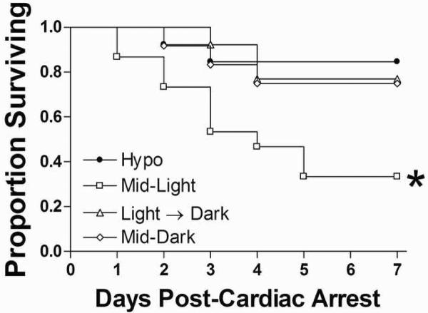
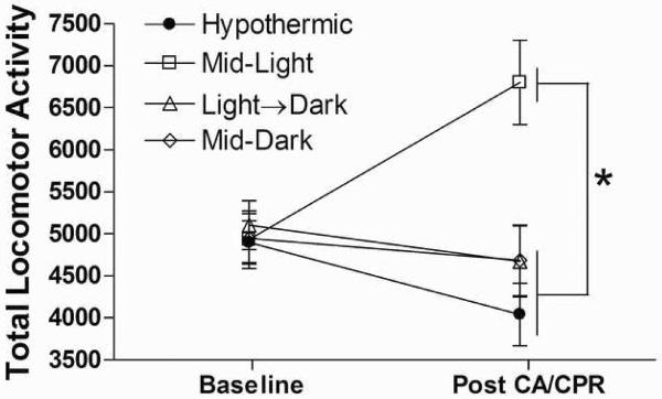
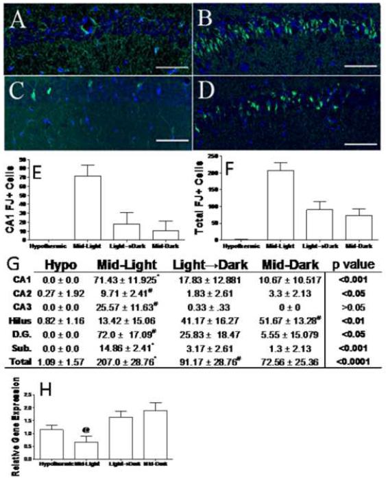
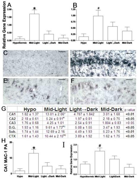
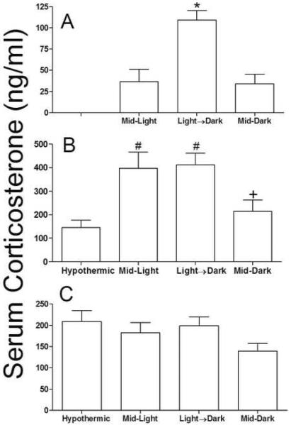
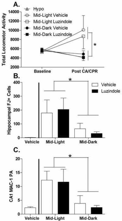
Ischemic events in humans are not evenly distributed across the day. To discriminate between temporal differences in the incidence of ischemia and susceptibility to ischemic events, we examined the outcome of global ischemia in a murine model at three time points during the day. Global cerebral ischemia in mice during the light phase impairs survival and exacerbates outcome compared to ischemia at other times of the day. Specifically, mice that underwent cardiac arrest during the light phase had greater numbers of degenerating neurons, greater microglial activation, and increased proinflammatory cytokine production in the ischemia-vulnerable hippocampus, as well as increased locomotor activity. Time-of-day differences were not altered by the melatonin receptor antagonist luzindole. Our results document that brain tissue displays endogenous fluctuations in susceptibility to ischemic damage and demonstrate that small differences in time of onset can significantly influence ischemic outcomes.


Figures

Figure 1
Figure 1. Time-of-day determines cardiac arrest survival
Cardiac arrest reduced survival in normothermic mice that underwent CA/CPR during the Mid-Light period. Eighty-four percent of mice in the hypothermic groups survived CA/CPR. Among the normothermic groups, mice in the Light→Dark transition group and the Mid-Dark group survived at 76.9% and 75%, respectively. In contrast, only 33% of mice that underwent CA/CPR during the light period (Mid-Light group) survived to day 7 post-reperfusion. Thus, timing of CA/CPR significantly altered survival (Kaplan-Meier Survival Analysis; χ2=10.19, p<0.05). *Significantly different from all other groups at p<0.05.
Figure 2
Figure 2. Time-of-day determines the behavioral consequences of cardiac arrest
Cardiac arrest-induced behavioral hyperactivity was observed only in mice that underwent cerebral ischemia during the Mid-Light period. * Mid-Light differs from all other groups post CA/CPR (p<0.05; data are presented as mean ±SEM). n=6–10/group.
Figure 3
Figure 3. Time-of-day determines that cell death responses to ischemia
Representative sections of Fluoro-jade C (degenerating neurons; green) and DAPI (nuclear counterstain; blue) stained tissue from the CA1 region of mice that underwent hypothermic CA/CPR (a) or normothermic CA/CPR during the Mid-Light (b), Light-Dark transition (c), or Mid-Dark period (d). Quantification of Fluoro Jade positive neurons in the CA1 field (e) and across the whole hippocampus (f). Table of mean (±SEM) for Fluoro-jade positive neurons in selected regions (g). Hippocampal bcl-2 gene expression relative to 18s rRNA gene expression at 24 h post-CA/CPR (h). Scale bar =100μm. CA -Cornu Ammonis, D.G. dentate gyrus, Sub. subiculum. * indicates significantly different from all other groups at p<0.05; # indicates significantly different from hypothermic mice at p<0.05. n=7–11/group for histology and 4–10/group for PCR.
Figure 4
Figure 4. Time-of-day determines inflammatory responses to cardiac arrest
Proinflammatory cytokine gene expression was potentiated by CA/CPR during the Mid-Light. (a) Il-1β and (b) Tnfα mRNA gene expression relative to 18s rRNA. Microglial activation was also potentiated in mice that underwent ischemia during the middle of the light period. Representative sections of MAC-1 stained sections through the CA1 field of the hippocampus in mice that underwent hypothermic CA/CPR (c) or normothermic CA/CPR in the Mid-Light (d), Light→ Dark Transition (e) or in the Mid-Dark (f). Table of proportional areas of microglial activation across the hippocampus and associated regions (g). (h) MAC-1 proportional area in the CA1 field. (i) Immunohistochemical evidence of increased microglial activation was confirmed with MAC-1 rat-PCR 24 h post-CA/CPR. Data are presented as the mean ±SEM. * significantly different from all other groups (p<0.05); # significantly different from hypothermic mice (p<0.05); c significantly different from mid-dark mice (P<0.05); d significantly different from Light→Dark mice (p<0.05). Scale bar =100μm. CA-Cornu Ammonis, D.G. dentate gyrus, Sub. subiculum, CTX, posterior parietal cortex n=7–11/group for histology and 4– 10/group for PCR.
Figure 5
Figure 5. Circulating glucocorticoids may not explain time-of-day differences in ischemic outcome
Serum corticosterone (a) 24 h prior to CA/CPR, (b) 24 h post-reperfusion or (c) seven days post-reperfusion. Data are presented as the mean ±SEM. *Significantly different from all other groups (P<0.05). #Significantly different from hypothermic mice (p<0.05). + Significantly different than Light→Dark (p<0.05). n= 4–10/group.
Figure 6
Figure 6. Melatonin receptor antagonists do not alter time-of-day differences in ischemic outcome
Treatment with luzindole 30 minutes prior to CA/CPR did not alter time of day differences in (A) post-ischemic hyperactivity (B), or time-of-day differences in FJ+ cells throughout the hippocampus (C) and microglial activation in the CA1 field. p> 0.05 in each comparison between luzindole and vehicle. Data are presented as the mean ± SEM. FJ- Fluoro Jade. *indicates significantly differences between the Mid-Light and Mid-dark groups. n=7–10/group.

Similar articles

Cited by

References

    1. Abe M, et al. Circadian rhythms in isolated brain regions. J Neurosci. 2002;22:350–6. - PMC - PubMed
    1. Antolin I, et al. Neurohormone melatonin prevents cell damage: effect on gene expression for antioxidant enzymes. Faseb J. 1996;10:882–90. - PubMed
    1. Bain RJ, et al. Serum cortisol levels predict infarct size and patient mortality. International Journal of Cardiology. 1992;37:145–50. - PubMed
    1. Barlow-Walden LR, et al. Melatonin stimulates brain glutathione peroxidase activity. Neurochemistry International. 1995;26:497–502. - PubMed
    1. Becker LB, et al. Outcome of CPR in a large metropolitan area--where are the survivors? Annals of Emergency Medicine. 1991;20:355–61. - PubMed

Publication types