. 2009 Oct;30(30):5910-7.
doi: 10.1016/j.biomaterials.2009.06.034. Epub 2009 Aug 6.

Scaffold-free vascular tissue engineering using bioprinting

Affiliations

Scaffold-free vascular tissue engineering using bioprinting

Cyrille Norotte et al. Biomaterials. 2009 Oct.

Abstract

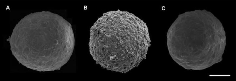
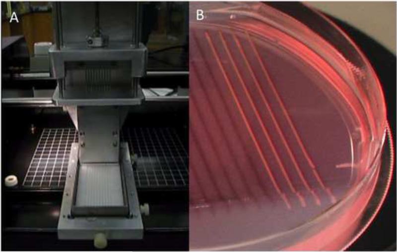
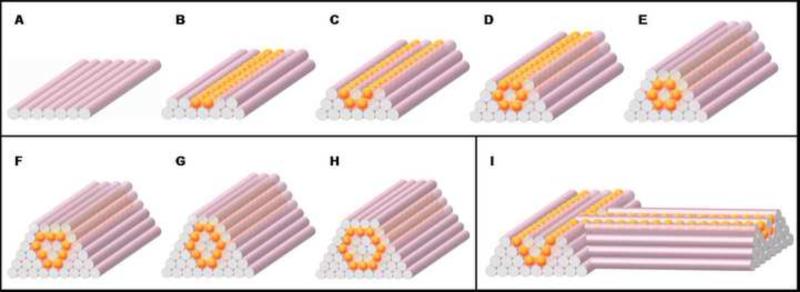
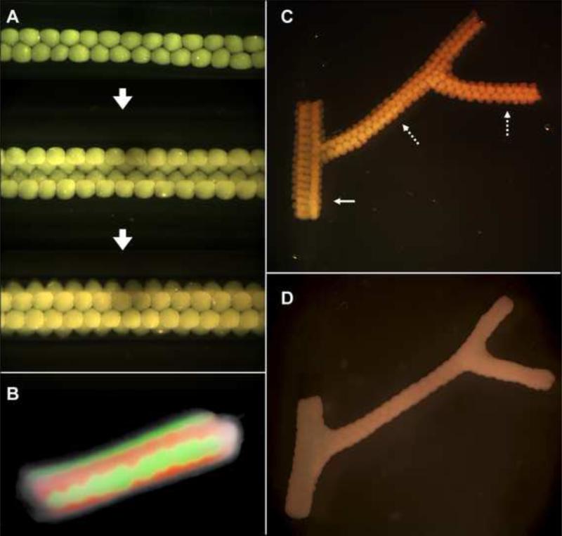
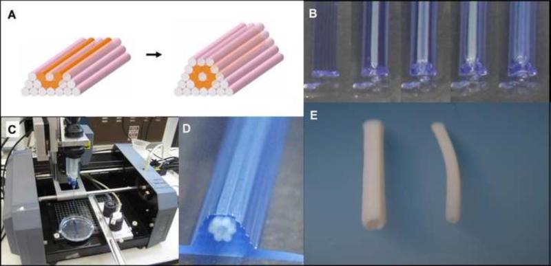
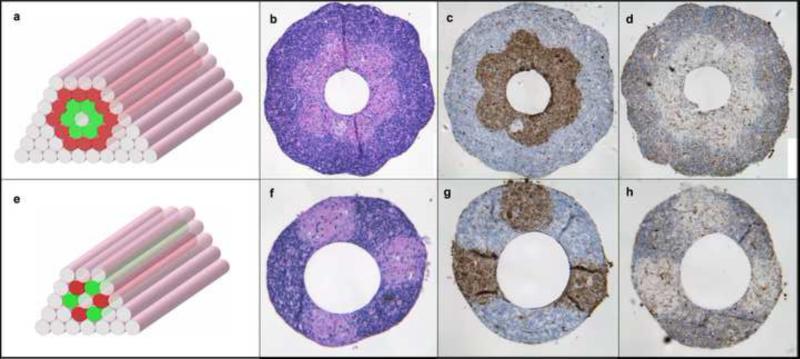
Current limitations of exogenous scaffolds or extracellular matrix based materials have underlined the need for alternative tissue-engineering solutions. Scaffolds may elicit adverse host responses and interfere with direct cell-cell interaction, as well as assembly and alignment of cell-produced ECM. Thus, fabrication techniques for production of scaffold-free engineered tissue constructs have recently emerged. Here we report on a fully biological self-assembly approach, which we implement through a rapid prototyping bioprinting method for scaffold-free small diameter vascular reconstruction. Various vascular cell types, including smooth muscle cells and fibroblasts, were aggregated into discrete units, either multicellular spheroids or cylinders of controllable diameter (300-500 microm). These were printed layer-by-layer concomitantly with agarose rods, used here as a molding template. The post-printing fusion of the discrete units resulted in single- and double-layered small diameter vascular tubes (OD ranging from 0.9 to 2.5mm). A unique aspect of the method is the ability to engineer vessels of distinct shapes and hierarchical trees that combine tubes of distinct diameters. The technique is quick and easily scalable.


Figures

Fig. 1
Fig. 1
Scanning electron micrographs of 300 μm diameter multicellular spheroids of HUSMCs (A), CHO cells (B) and HFBs (C) employed in the present study. HUSMC and HSF spheroids display similar morphology with smooth and uniform surface and contain respectively about 8,000 and 15,000 cells. In contrast, CHO spheroids assume a berry-like shape suggesting that surface cells adhere more weakly to inner cell layers. Scale bar: 100 μm.
Fig. 2
Fig. 2
Preparation of cylindrical bioink. A. Upon cell aggregation in micropipettes, ten multicellular cylinders were simultaneously extruded using a special-purpose attachment to a specifically designed bioprinter. B. Non-adhesive agarose mold in which extruded cylinders were matured prior to be used as bioink.
Fig. 3
Fig. 3
Design template for tubular structures. (A-E) Deposition scheme for the smallest diameter tube that can be built of agarose rods (pink) and multicellular spheroids (orange) of the same diameter. (F-H) More complex tubular structures. (I) Scheme for a branching structure.
Fig. 4
Fig. 4
Fusion patterns of multicellular spheroids assembled into tubular structures. A. HSF spheroids assembled according to template in Fig. 3A-E. B. Fusion pattern after 7 days of a tube assembled from fluorescently labeled red and green sequences of CHO spheroids. C. Branched structure built of 300 μm HSF spheroids with branches of 1.2 mm (solid arrow) and 0.9 mm (broken arrows). D. The fused branched construct after 6 days of deposition.
Fig. 5
Fig. 5
Bioprinting tubular structures with cellular cylinders. A. Design template analogous to the one in Fig. 3. B. Layer-by-layer deposition of agarose cylinders (stained here in blue for better visualization) and multicellular pig SMC cylinders. C. The bioprinter (see Materials and Methods) outfitted with two vertically moving print heads. D. The printed construct. E. Engineered pig SMC tubes of distinct diameters resulted after 3 days of post-printed fusion (left: 2.5 mm OD; right: 1.5 mm OD).
Fig. 6
Fig. 6
Building a double-layered vascular wall. A, B. HUVSMC and HSF multicellular cylinders were assembled according to specific patterns (HUVSMC: green; HSF: red) C-H. Histological examination of the structures after 3 days of fusion: H&E (C, F), smooth muscle α-actin (brown; D, G) and Caspase-3 (brown; E, H) stainings are shown. Note that the more complex construct in the upper row requires more time to fuse.
Fig. 7
Fig. 7
Maturation of bioprinted tubes composed of porcine aortic smooth muscle cells (left) in a perfusion bioreactor (right).

References

    1. Atala A, Bauer SB, Soker S, Yoo JJ, Retik AB. Tissue-engineered autologous bladders for patients needing cystoplasty. Lancet. 2006;367:1241–1246. - PubMed
    1. Kerker JT, Leo AJ, Sgaglione NA. Cartilage repair: synthetics and scaffolds: basic science, surgical techniques, and clinical outcomes. Sports Med Arthrosc. 2008;16:208–216. - PubMed
    1. Priya SG, Jungvid H, Kumar A. Skin tissue engineering for tissue repair and regeneration. Tissue Eng Part B Rev. 2008;14:105–218. - PubMed
    1. Shin'oka T, Imai Y, Ikada Y. Transplantation of a tissue-engineered pulmonary artery. N Engl J Med. 2001;344:532–533. - PubMed
    1. van Tienen TG, Hannink G, Buma P. Meniscus replacement using synthetic materials. Clin Sports Med. 2009;28:143–156. - PubMed

Publication types

Substances