. 2009 Sep 29;214(1-2):67-77.
doi: 10.1016/j.jneuroim.2009.06.023. Epub 2009 Aug 6.

Beneficial role of the GPR30 agonist G-1 in an animal model of multiple sclerosis

Affiliations

Beneficial role of the GPR30 agonist G-1 in an animal model of multiple sclerosis

Eric Blasko et al. J Neuroimmunol. .

Abstract

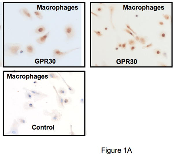
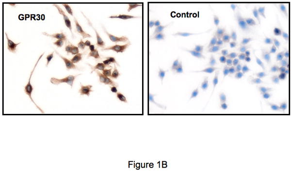
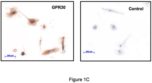
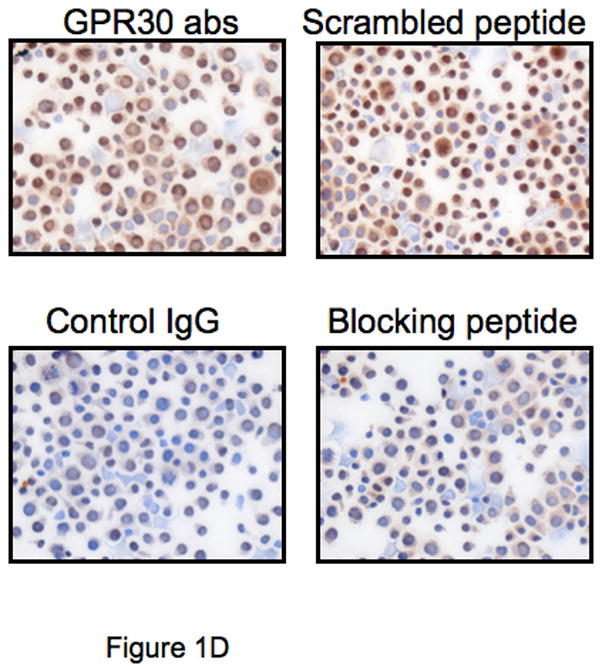
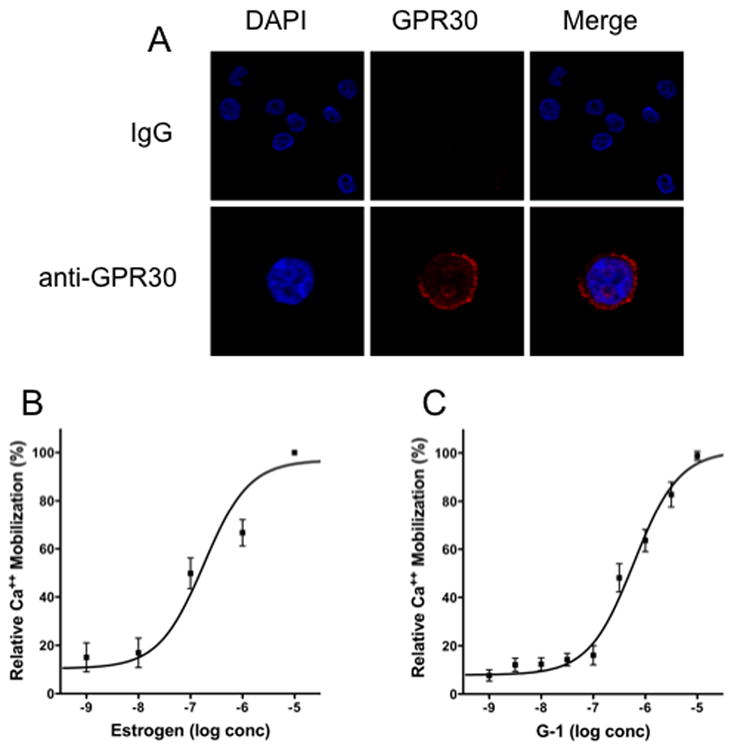
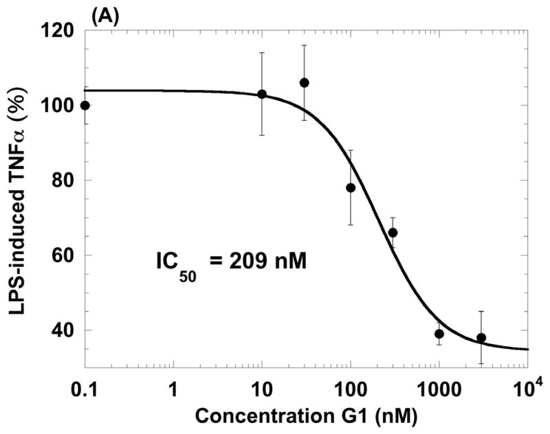
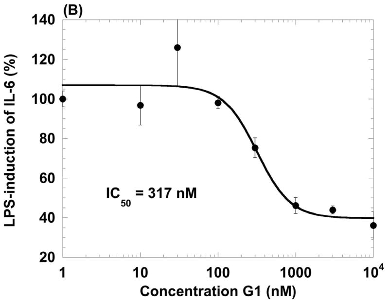
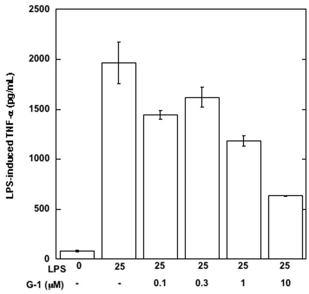
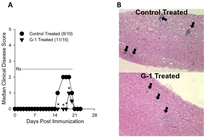
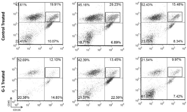
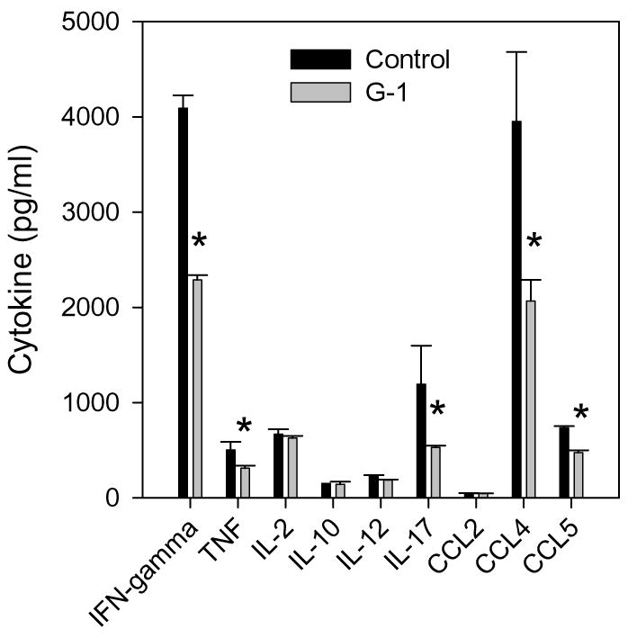
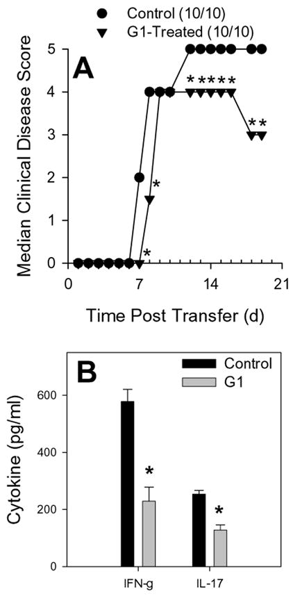
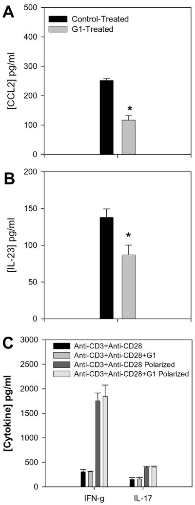
The beneficial effects of estrogens in multiple sclerosis are thought to be mediated exclusively by the classical nuclear estrogen receptors ERalpha and ERbeta. However, recently many reports revealed that estrogens are able to mediate rapid signals through a G protein-coupled receptor (GPCR), known as GPR30. In the present study, we set out to explore whether effects mediated through this receptor were anti-inflammatory and could account for some of the beneficial effects of estrogen. We demonstrate that GPR30 is expressed in both human and mouse immune cells. Furthermore a GPR30-selective agonist, G-1, previously described by us, inhibits the production of lipopolysaccharide (LPS)-induced cytokines such as TNF-alpha and IL-6 in a dose-dependent manner in human primary macrophages and in a murine macrophage cell line. These effects are likely mediated solely through the estrogen-specific receptor GPR30 since the agonist G-1 displayed an IC(50) far greater than 10 microM on the classical nuclear estrogen receptors as well as a panel of 25 other GPCRs. Finally, we show that the agonist G-1 is able to reduce the severity of disease in both active and passive EAE models of multiple sclerosis in SJL mice and that this effect is concomitant with a G-1-mediated decrease in proinflammatory cytokines, including IFN-gamma and IL-17, in immune cells harvested from these mice. The effect of G-1 appears indirect, as the GPR30 agonist did not directly influence IFN-gamma or IL-17 production by purified T cells. These data indicate that G-1 may represent a novel therapeutic agent for the treatment of chronic autoimmune, inflammatory diseases.


Figures

Figure 1
Figure 1
GPR30 expression by immunohistochemical staining (brown) in primary immune cells and cell lines. (A) Human macrophages, (B) Mouse RAW 264.7 cells, (C) Rat microglia and (D) Human regulatory T cells all stain with an antibody against GPR30. Staining is primarily cytoplasmic in the mouse and rat cells, while in the human macrophages and T cells it is both cytoplasmic and nuclear. There is little to no staining with a control antibody. In addition, staining is abolished by preincubation with a C-terminal peptide to GPR30 but not with a scrambled version of the same peptide (D). Nuclei are lightly counterstained with hematoxylin. All images taken with a 63- oil objective on a Zeiss Axioskop. Scale bars = 100 μm in D.
Figure 1
Figure 1
GPR30 expression by immunohistochemical staining (brown) in primary immune cells and cell lines. (A) Human macrophages, (B) Mouse RAW 264.7 cells, (C) Rat microglia and (D) Human regulatory T cells all stain with an antibody against GPR30. Staining is primarily cytoplasmic in the mouse and rat cells, while in the human macrophages and T cells it is both cytoplasmic and nuclear. There is little to no staining with a control antibody. In addition, staining is abolished by preincubation with a C-terminal peptide to GPR30 but not with a scrambled version of the same peptide (D). Nuclei are lightly counterstained with hematoxylin. All images taken with a 63- oil objective on a Zeiss Axioskop. Scale bars = 100 μm in D.
Figure 2
Figure 2
Expression and function of GPR30 in human promyelocytic HL60 cells. (A) HL60 cells were cytospun onto coated slides and stained with either IgG or GPR30 immune serum (red) and nuclei were counterstained with DAPI (blue). Calcium mobilization in indo-1-loaded HL60 cells was determined in response to estrogen (B) and the GPR30 agonist G-1 (C).
Figure 3
Figure 3
Agonist properties of the GPR30 agonist G-1 on human primary macrophages. G-1 inhibits LPS-induced production of TNF-α (A) and IL-6 (B) in human macrophages in a dose-dependent manner. Data values are mean +/− S.D. of triplicate points within the same experiment; the data are representative of three separate experiments
Figure 3
Figure 3
Agonist properties of the GPR30 agonist G-1 on human primary macrophages. G-1 inhibits LPS-induced production of TNF-α (A) and IL-6 (B) in human macrophages in a dose-dependent manner. Data values are mean +/− S.D. of triplicate points within the same experiment; the data are representative of three separate experiments
Figure 4
Figure 4
Agonist properties of the GPR30 agonist G-1 on the mouse macrophage cell line RAW 264.7 G-1 inhibits LPS-induced TNF-α in RAW 264.7 cells in a dose-dependent manner. Data values are mean +/− S.D. of triplicate points within the same experiment; the data are representative of four separate experiments
Figure 5
Figure 5
GPR30 agonist treatment reduced the severity of actively induced experimental autoimmune encephalomyelitis. A) SJL mice (5–7 weeks old) were immunized s.c. with 50 μg PLP139-151 and CFA (400 μg Mycobacterium tuberculosis). Mice were treated with 50 mg/kg/day G-1 daily for 21 days beginning at the day of disease induction (Rx). Control mice were similarly treated with vehicle (5% DMSO, 95% PEG-300). Clinical disease was scored as 0, asymptomatic; 1, tail atonia; 2, hind limb paresis; 2, unilateral hind limb paralysis; 4, bilateral hind limb paralysis; and 5, death. The data are shown as the median clinical disease score as a function of days post immunization. Disease incidence for each treatment group is indicated in parentheses. The G-1-treated group showed significantly decreased acute clinical disease score, but not incidence, compared to control mice (*, p<0.05). B) The CNS of representative mice from each treatment group was evaluated for histologic EAE by standard H&E staining. The photomicrographs (100x magnification) indicate less severe mononuclear cell lesions in the G-1- compared to control-treated mice (heavy black arrows). These data are representative of three similar, independent experimental replicates.
Figure 6
Figure 6
GPR30 agonist treatment reduced CNS macrophage accumulation. SJL mice (5–7 weeks old) were immunized s.c. with 50 μg PLP139-151 and CFA (400 μg Mycobacterium tuberculosis). Mice were treated with 50 mg/kg/day G-1 daily for 21 days beginning at the day of disease induction. Control mice were similarly treated with vehicle (5% DMSO, 95% PEG-300). When the control mice showed peak clinical disease (Figure 5), CNS was harvested from three representative mice in each of the control (disease scores=3, 3, 2) and G-1-treated (disease scores=1, 1, 1) groups. Leukocyte subpopulations were examined in each individual mouse by flow cytometry. The data show a decrease in CD45hiCD11b+ macrophage percentages in the CNS of G-1-treated mice. The data are representative of two similar, independent experimental replicates.
Figure 7
Figure 7
GPR30 agonist treatment inhibits inflammatory cytokine expression. Splenocytes from the individual mice analyzed in Figure 6 were re-stimulated with 50 μg/ml PLP139-151 peptide in vitro. Culture supernatants were harvested after 48 h and assessed for the presence of cytokines using Beadlyte Mouse Multi-cytokine Flex Kit assay. The data are shown as mean cytokine production (pg/ml from three individual mice ±SD) in cultures from G-1-treated mice compared to cells from control-treated mice. The results indicate significant (*, p<0.05) reductions in IFN-γ, TNF, IL-12, IL-17, CCL4, and CCL5 expression. The data are representative of two similar, independent experimental replicates.
Figure 8
Figure 8
GPR30 agonist treatment reduced severity of adoptively transferred experimental autoimmune encephalomyelitis. SJL mice (5–7 weeks old) were immunized s.c. with 50 μg PLP139-151 and CFA (400 μg Mycobacterium tuberculosis). Draining lymph node cells were harvested after 7 days and re-stimulated with 50 μg/mL PLP139-151 in the presence of 3 μM G-1 agonist or control DMEM medium with 5% FCS and 0.1% DMSO. Antigen-activated T cells were harvested after 72 hours of culture and 6×10⁶ blasts were transferred i.v. to naïve SJL recipient mice. Clinical disease was scored as 0, asymptomatic; 1, tail atonia; 2, hind limb paresis; 2, unilateral hind limb paralysis; 4, bilateral hind limb paralysis; and 5, death. A) The data are shown as the median clinical disease score as a function of days post lymphocyte transfer. The group that received G-1-treated cells showed significantly decreased clinical disease compared to recipients of control-treated cells (Mann-Whitney *, p<0.05). B) Lymphocytes were harvested from the CNS of representative recipient mice, restimulated with specific antigen and irradiated splenic APC, and the resulting culture supernatants were assessed for IFN-γ and IL-17 by ELISA. The results indicate that antigen-specific T cells recovered from the CNS of mice that received G-1-treated lymphocytes produced significantly less (*, p<0.05) IFN-γ and IL-17. The results are representative of two independent replicate experiments.
Figure 9
Figure 9
GPR30 agonist treatment reduced CNS cytokine expression. SJL mice (5–7 weeks old) were immunized s.c. with 50 μg PLP139-151 and CFA (400 μg Mycobacterium tuberculosis). Draining lymph node cells were harvested after 7 days and re-stimulated with 50 μg/mL PLP139-151 in the presence of 3 μM G-1 agonist or control DMEM medium with 5% FCS and 0.1% DMSO. When the control group showed peak clinical disease, three representative mice from each group were harvested and the CNS tissue was analyzed for the presence of CCL2 (A) and IL-23 (B) by ELISA. The group that received G-1-treated cells showed significantly decreased CNS CCL2 and IL-23 compared to recipients of control-treated cells (*, p<0.05). C) Purified CD4+ T cells were stimulated in vitro with immobilized anti-CD3 and anti-CD28 in the presence or absence of 3 μM G-1 under normal and polarizing conditions. Polarizing conditions included IL-12 and anti-IL-4 for Th1 (IFN-γ) and TGF-β, IL-6 and IL-23 for Th17 (IL-17). The data indicate no effect of G-1 on T cell cytokine secretion.

References

    1. Abramsky O. Pregnancy and multiple sclerosis. Ann Neurol. 1994;36(Suppl):S38–41. - PubMed
    1. Alderson KL, Zhou Q, Berner V, Wilkins DE, Weiss JM, Blazar BR, Welniak LA, Wiltrout RH, Redelman D, Murphy WJ. Regulatory and conventional CD4+ T cells show differential effects correlating with PD-1 and B7-H1 expression after immunotherapy. J Immunol. 2008;180:2981–2988. - PMC - PubMed
    1. Arnason BG, Richman DP. Effects of estrogen, progestin and combined estrogen-progestin oral contraceptive preparations on experimental allergic encephalomyelitis. Trans Am Neurol Assoc. 1969;94:54–58. - PubMed
    1. Bologa CG, Revankar CM, Young SM, Edwards BS, Arterburn JB, Kiselyov AS, Parker MA, Tkachenko SE, Savchuck NP, Sklar LA, Oprea TI, Prossnitz ER. Virtual and biomolecular screening converge on a selective agonist for GPR30. Nat Chem Biol. 2006;2:207–212. - PubMed
    1. Carmeci C, Thompson DA, Ring HZ, Francke U, Weigel RJ. Identification of a gene (GPR30) with homology to the G-protein-coupled receptor superfamily associated with estrogen receptor expression in breast cancer. Genomics. 1997;45:607–617. - PubMed

MeSH terms

Substances