

Chronic inflammatory and non-inflammatory diseases of the gastrointestinal tract in cats: diagnostic advantages of full-thickness intestinal and extraintestinal biopsies
- PMID: 19664949
- PMCID: PMC10911445
- DOI: 10.1016/j.jfms.2009.07.004
Chronic inflammatory and non-inflammatory diseases of the gastrointestinal tract in cats: diagnostic advantages of full-thickness intestinal and extraintestinal biopsies
Abstract
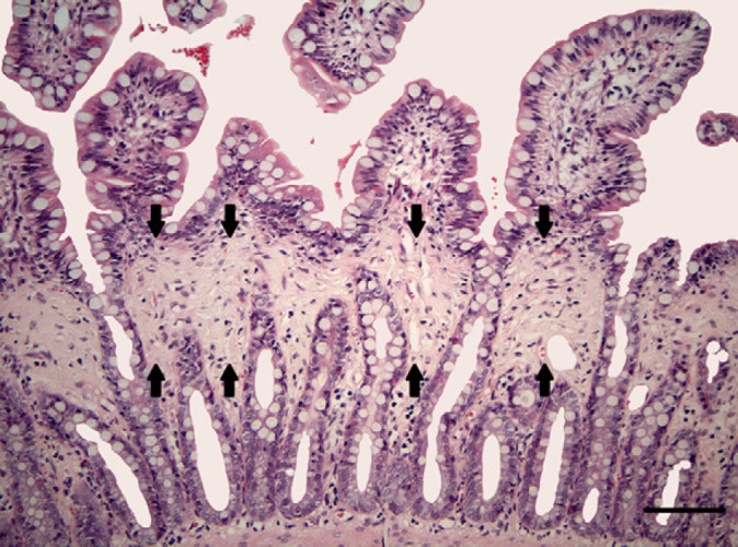
An evaluation of histological findings in full-thickness biopsies from the gastrointestinal tract (GIT) and extraintestinal samples of 43 cats with chronic GIT disease signs was performed. In the majority of cases (46.5%) inflammatory bowel disease, ie, lymphocytic-plasmacytic enteritis/colitis (32.6%), eosinophilic gastroenterocolitis (11.6%) and mixed inflammatory infiltration (2.3%), was diagnosed. Furthermore, in four animals non-inflammatory mucosal band-shaped fibrosis (9.3%), and in 10 cats (23.3%) a diffuse lymphoma, was found. Six cats displayed only a gastritis (7.0%) or lymphangiectasia (7.0%), respectively. In two cats a mast cell tumour (4.7%) was diagnosed. In one cat no histopathological lesions were found. The availability of transmural biopsies from all segments of the intestine and the collection of extraintestinal samples, especially mesenteric lymph nodes, is especially helpful for diagnosing intestinal tumours such as lymphomas and tumours of mast cell origin.
Copyright 2009 ESFM and AAFP. Published by Elsevier Ltd. All rights reserved.
Figures


Similar articles
-
Retrospective study on the diagnostic value of full-thickness biopsies from the stomach and intestines of dogs with chronic gastrointestinal disease symptoms.Vet Pathol. 2006 Nov;43(6):1000-3. doi: 10.1354/vp.43-6-1000. Vet Pathol. 2006. PMID: 17099159
-
The diagnostic relevance of mesenteric lymph node biopsy in small intestinal lymphoma in cats.J Vet Intern Med. 2024 Jul-Aug;38(4):2316-2323. doi: 10.1111/jvim.17095. Epub 2024 Jun 10. J Vet Intern Med. 2024. PMID: 38858174 Free PMC article.
-
Utility of endoscopic biopsies of the duodenum and ileum for diagnosis of inflammatory bowel disease and small cell lymphoma in cats.J Vet Intern Med. 2011 Nov-Dec;25(6):1253-7. doi: 10.1111/j.1939-1676.2011.00831.x. Epub 2011 Nov 1. J Vet Intern Med. 2011. PMID: 22092613
-
Alimentary neoplasia in geriatric dogs and cats.Vet Clin North Am Small Anim Pract. 2012 Jul;42(4):693-706, vi. doi: 10.1016/j.cvsm.2012.04.006. Epub 2012 May 10. Vet Clin North Am Small Anim Pract. 2012. PMID: 22720809 Review.
-
Ultrasonography of small intestinal inflammatory and neoplastic diseases in dogs and cats.Vet Clin North Am Small Anim Pract. 2011 Mar;41(2):329-44. doi: 10.1016/j.cvsm.2011.01.002. Epub 2011 Mar 3. Vet Clin North Am Small Anim Pract. 2011. PMID: 21486639 Review.
Cited by
-
Feline low-grade alimentary lymphoma: an emerging entity and a potential animal model for human disease.BMC Vet Res. 2018 Oct 11;14(1):306. doi: 10.1186/s12917-018-1635-5. BMC Vet Res. 2018. PMID: 30305106 Free PMC article. Review.
-
Endoscopic Biopsies and Histopathological Findings in Diagnosing Chronic Gastrointestinal Disorders in Dogs and Cats.Vet Med Int. 2020 Oct 9;2020:8827538. doi: 10.1155/2020/8827538. eCollection 2020. Vet Med Int. 2020. PMID: 33133490 Free PMC article. Review.
-
Mechanisms, causes, investigation and management of vomiting disorders in cats: a literature review.J Feline Med Surg. 2013 Apr;15(4):237-65. doi: 10.1177/1098612X12473466. Epub 2013 Feb 12. J Feline Med Surg. 2013. PMID: 23403690 Free PMC article. Review.
-
Ability of ultrasonography to predict the presence and location of histologic lesions in the small intestine of cats.J Vet Intern Med. 2019 May;33(3):1278-1285. doi: 10.1111/jvim.15471. Epub 2019 Mar 7. J Vet Intern Med. 2019. PMID: 30847975 Free PMC article.
-
Outbreak in African lions of Yersinia pseudotuberculosis infection, with aberrant bacterial morphology.J Vet Diagn Invest. 2022 Mar;34(2):334-338. doi: 10.1177/10406387211072822. Epub 2022 Jan 17. J Vet Diagn Invest. 2022. PMID: 35037547 Free PMC article.
References
-
- Jergens A.E., Moore F.M., Haynes J.S., Miles K.G. Idiopathic inflammatory bowel disease in dogs and cats: 84 cases (1987–1990), J Am Vet Med Assoc 201, 1992, 1603–1608. - PubMed
-
- Dennis J.S., Kruger J.M., Mullaney T.P. Lymphocytic/plasmacytic colitis in cats: 14 cases (1985–1990), J Am Vet Med Assoc 202, 1993, 313–318. - PubMed
-
- Guilford W.G. Idiopathic inflammatory bowel disease. Guilford W.G., Center S.A., Strombeck D.R., Williams D.A., Meyer D.J. Strombeck's small animal gastroenterology, 1996, WB Saunders: Philadelphia, 451–486.
-
- Tams T.R. Chronic feline inflammatory bowel disorders. Part I. Idiopathic inflammatory bowel disease, Compend Contin Educ Pract Vet 8, 1986, 371–376.
-
- Jergens A.E. Feline idiopathic inflammatory bowel disease, Compend Contin Educ Pract Vet 14, 1992, 509–518.
MeSH terms
LinkOut - more resources
Full Text Sources
Medical
Miscellaneous
