. 2009 Sep 4;5(3):267-78.
doi: 10.1016/j.stem.2009.06.004. Epub 2009 Aug 6.

Distinct self-renewal and differentiation phases in the niche of infrequently dividing hair follicle stem cells

Affiliations

Distinct self-renewal and differentiation phases in the niche of infrequently dividing hair follicle stem cells

Ying V Zhang et al. Cell Stem Cell. .

Abstract

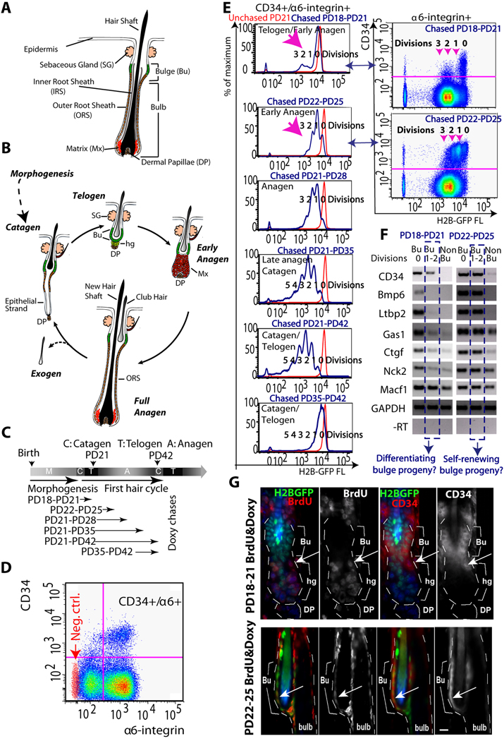
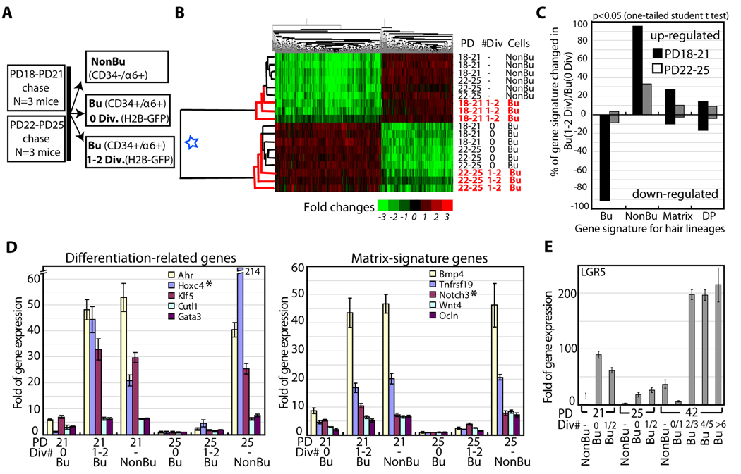
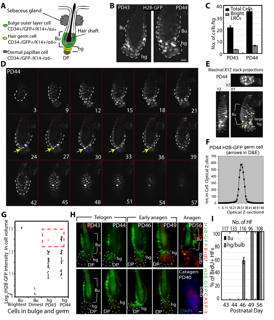
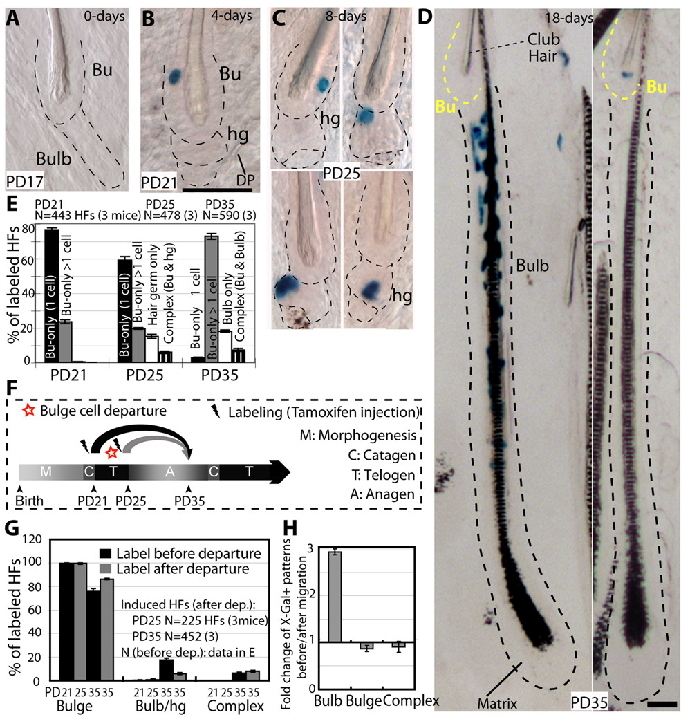
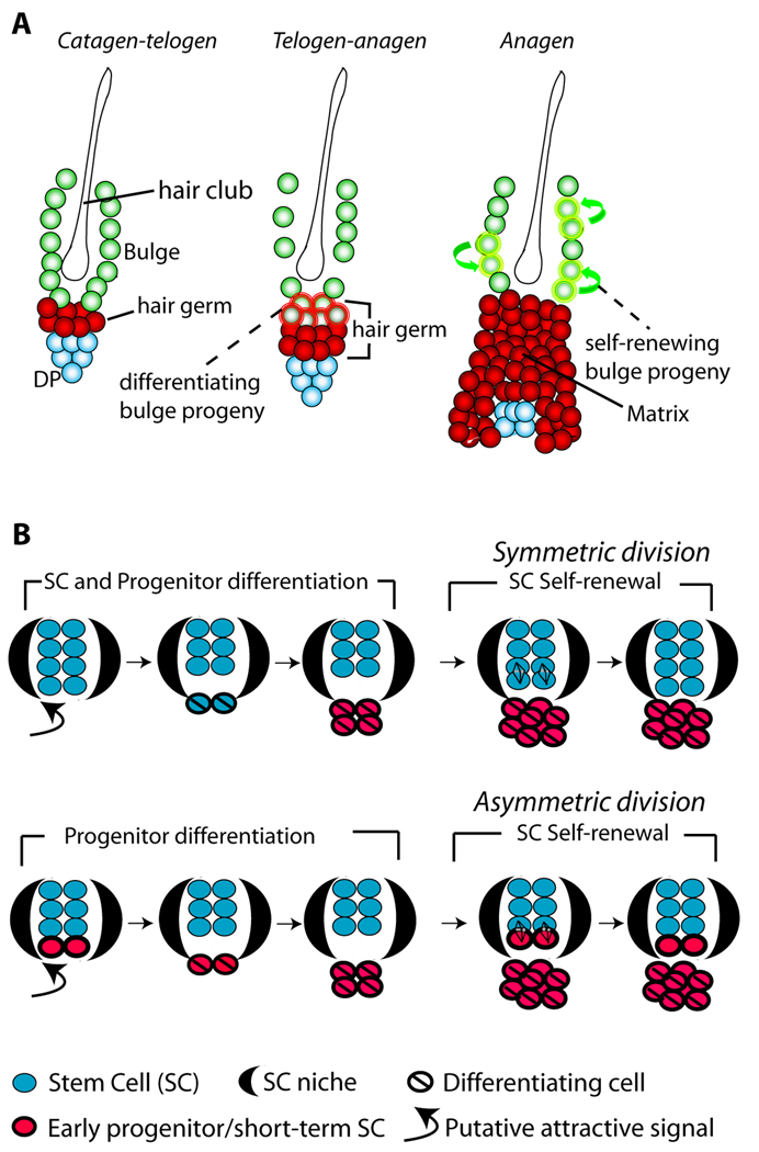
In homeostasis of adult vertebrate tissues, stem cells are thought to self-renew by infrequent and asymmetric divisions that generate another stem cell daughter and a progenitor daughter cell committed to differentiate. This model is based largely on in vivo invertebrate or in vitro mammal studies. Here, we examine the dynamic behavior of adult hair follicle stem cells in their normal setting by employing mice with repressible H2B-GFP expression to track cell divisions and Cre-inducible mice to perform long-term single-cell lineage tracing. We provide direct evidence for the infrequent stem cell division model in intact tissue. Moreover, we find that differentiation of progenitor cells occurs at different times and tissue locations than self-renewal of stem cells. Distinct fates of differentiation or self-renewal are assigned to individual cells in a temporal-spatial manner. We propose that large clusters of tissue stem cells behave as populations whose maintenance involves unidirectional daughter-cell-fate decisions.


Figures

Figure 1
Figure 1
Distinct characteristics of bulge cells newly generated at different hair cycle stages. (A) The hair follicle structure. (B) Hair cycle phases of growth (anagen), destruction (catagen) and quiescence (telogen) (see text). (C) Doxycycline (doxy) chases in 1st (synchronous) hair cycle to document cell proliferation quantitatively. PD, postnatal day. (D) FACS dot plot of live (PI negative) skin cells stained for CD34 and α6-integrin surface expression (PD21-PD28 chase). Red dots, secondary antibody alone for CD34 staining. (E) FACS histograms of CD34+/α6+ bulge cells from individual mice at stages indicated. Note distinct proliferation profiles at different stages (pink arrows). (F) Cells were sorted and analyzed by RT-PCR for expression of bulge-preferred factors (Tumbar et al, 2004). (G) Skin images from 3-day BrdU labeled and doxy treated mice showed BrdU staining in hair germ (hg) at PD18-PD21 and in bulge (Bu) at PD22-PD25. Arrows indicate CD34+/BrdU+ proliferating cells. Note that lower CD34 levels at the bulge/germ transition zone at PD18- PD21. All experiments were repeated in ≥ 3 mice per stage. DP, dermal papillae. Scale bar in G, 10µm.
Figure 2
Figure 2
Expression profiling at 2-hair cycle stages. (A) Cell subpopulations sorted and profiled by expression microarrays. (B) Gene (top), and cell population (left) dendogram heat map shows 2 main population clusters (blue star). Newly divided CD34+/α6+ cells (Bu 1–2 Div) cluster with a different group at each stage. (C) Genes changed in Bu (1–2 Div) cell populations relative to Bu (0 Div) are shown as percent of 4 hair lineages gene signatures: CD34+/α6+ bulge (Bu). CD34−/α6+ non-bulge (NonBu), matrix (Mx), dermal papillae (DP) (Table S2). Note down-regulation of bulge signature genes and higher up-regulation of Mx genes at PD18-PD21. DP was a negative control as an unrelated lineage and showed low and similar representation in both populations. (D). QRT-PCR (relative to GAPDH) of several Non-Bu or Matrix signature genes found up-regulated in our microarray in Bu (1–2 Div) relative to Bu (0 Div) shown with SEM. N=2 mice per stage (triplicate wells). Regular RT-PCR performed for 4 additional mice per stage is not shown. (E) Same QRT-PCR analysis for LGR5; a second experiment with another set of mice at each stage is shown in Fig. S2.
Figure 3
Figure 3
Bulge-cell departure from the niche during the quiescent phase. (A) Schematic of telogen HF structure, showing 2 layers of cells surrounding the shaft that make the bulge (Bu), and the hair germ (hg) underneath. DP, dermal papillae. (B) Confocal image from whole HFs in 100µm skin sections of pTRE H2B-GFP/K5tTA mice with doxy chase from PD21. Note increased number of hg cells by PD44, quantified in (C) and shown with SEM (N=39 HFs). (D) Confocal stack images of whole HF collected side by side from skin sections of pTRE-H2B-GFP/K5tTA mice at PD43 and PD44 (3-wks doxy chase; PD43 is shown in Fig. S3). Optical z-stacks are shown as tiled images. Numbers indicate actual optical slice and arrows point to a bright H2B-GFP cell throughout the stack located in hg at PD44, normally found in bulge at PD43. TOPRO-3 was used as DNA counterstain for revealing the hair follicle structures (not shown). (E) Stacks in (D) are shown as maximal projection through the slices on XY, ZY, and XZ plane. Arrow points to the same cells as in (D). (F) Total intensity after background subtraction (Int) in each optical z-section for cell indicated by arrow in (D&E) used to obtain total 3D intensity, which was then used to generate the plot in G. (G) H2B-GFP intensity/hg cell volume shows more bright cells at PD44 relative to PD43 (see Fig. S3); brightest (0 divisions) and dimmest bulge cells are shown for comparison. There are only 1–2 cells with 0 division, and they were localized in the bulge at PD43&PD44. (H) Skin section for mice fed doxy and BrdU for 3 days at stages indicated, and stained for BrdU (top) or caspase (bottom). (I) Frequencies of BrdU+ HFs (N at top) averaged in 3 mice/stage, with SEM. Scale bars, 10 µm.
Figure 4
Figure 4
Single-bulge cells lineage tracing during 1st adult hair cycle. (A–D) X-Gal stained skin sections (60–90µm) from K14CreER x Rosa26R mice tamoxifen injected at PD17 and sacrificed at times indicated. Dotted lines delineate the bulge (Bu) and hair germ (hg) or bulb compartments. Scale bar, 50 µm. (E) Frequencies of X-Gal patterns shown as average with SEM (N.3 mice/stage). DP, dermal papillae. (F) Scheme of chases PD17-PD21 or PD21-PD25, and X-Gal patterns in HFs at PD35 in anagen (G), re-shown as fold changes (H).
Figure 5
Figure 5
Multipotency of bulge cells in vivo. (A) Hair bulb with indicated differentiated lineages. Outer root sheath (ORS); IRS (inner root sheath made of cuticle (C), Huxley (Hu), and Henle (He) cell layers); hair shaft “core” made of 3 cell layers: (Cu) cuticle, (Co) cortex, (Me) medulla. Cp is ORS companion layer, included with the IRS category for simplification. (B) Quantification of PD35 labelling patterns in mice injected with tamoxifen at PD17. Lineages containing X-Gal+ cells are indicated at bottom, shown with SEM in 3 mice (>60 labelled HFs/mouse). (C&D) X-Gal bulb-only patterns in thick (90µm) or thin (20µm) serial sections. Scale bar, 50µm. (E) Cross-sectional images of X-gal stained skin from PD17-PD35 chase 20µm shows multiple differentiated layers in a majority of HFs. Scale bar, 10 µm. (F) Patterns of labelling, with SEM shown as % of a defined pattern from total labelled HFs (N= 3 mice, > 200 labelled HFs/mouse).
Figure 6
Figure 6
Long-term single-bulge cell lineage tracing. (A) Scheme of 1st telogen labelling and long-term chases. (B,E&H) Illustrative images from thick (60–90µm) skin sections show X-Gal staining in bulge (Bu) (yellow line) and bulb/germ (white line) compartments. Image in (E) shows a rare patch of frequent HF induction illustrating the coexistence of all 3 staining patterns in one skin region. Image in H shows bulb-only and bulge-only patterns after one year and at least 3 hair cycles post-labelling. The predominant patterns were complex (see counts in F). Scale bars, 50µm. (C&F) Quantification with SEM of X-Gal patterns at stages indicated. (D&G) Frequency of all labelled follicles counted at each stage (from data in Fig 4&Fig 6) show significant decline from one hair cycle to another.
Figure 7
Figure 7
Dynamics of hair follicle bulge stem cells during tissue homeostasis (A) The bulge cells undergo distinct phases of differentiation and self-renewal. First, at the telogen-anagen transition bulge cells depart the niche, proliferate and begin to differentiate to matrix progenitors of inner hair lineages. Second, bulge cells replenish their pool by divisions in anagen (self-renew), when newly generated bulge cells remain in the niche during the same hair cycle. (B) Models for adult SC renewal by illustrating symmetric (top) and asymmetric (bottom) divisions, and the predicted organization and dynamic of stem/progenitor cells in the niche in each scenario. Loss of SC identity may occur outside the niche (bulge) before or after the first differentiating division.

Similar articles

Cited by

References

    1. Barker N, van Es JH, Kuipers J, Kujala P, van den Born M, Cozijnsen M, Haegebarth A, Korving J, Begthel H, Peters PJ, Clevers H. Identification of stem cells in small intestine and colon by marker gene Lgr5. Nature. 2007;449:1003–1007. - PubMed
    1. Clayton E, Doupe DP, Klein AM, Winton DJ, Simons BD, Jones PH. A single type of progenitor cell maintains normal epidermis. Nature. 2007;446:185–189. - PubMed
    1. Cotsarelis G. Epithelial stem cells: a folliculocentric view. J Invest Dermatol. 2006;126:1459–1468. - PubMed
    1. Fuchs E. The tortoise and the hair: Slow-cycling cells in the stem cell race. Cell 137 811–819. 2009 - PMC - PubMed
    1. Greco V, Chen T, Rendl M, Schober M, Pasolli HA, Stokes N, Dela Cruz-Racelis J, Fuchs E. A two-step mechanism for stem cell activation during hair regeneration. Cell Stem Cell. 2009;4:155–169. - PMC - PubMed

Publication types

Associated data