. 2009 Oct 15;334(2):383-94.
doi: 10.1016/j.ydbio.2009.07.043. Epub 2009 Aug 6.

MAP kinase dependent cyclinE/cdk2 activity promotes DNA replication in early sea urchin embryos

Affiliations

MAP kinase dependent cyclinE/cdk2 activity promotes DNA replication in early sea urchin embryos

J Kisielewska et al. Dev Biol. .

Abstract

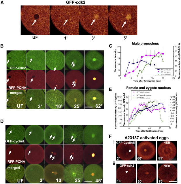
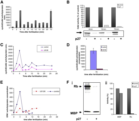
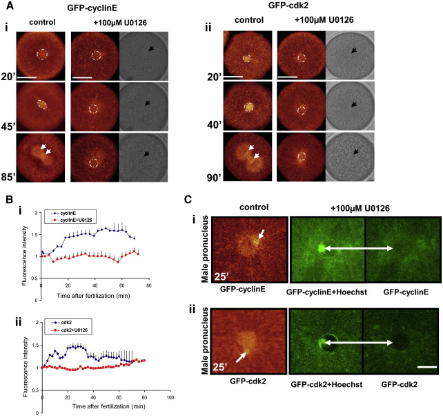
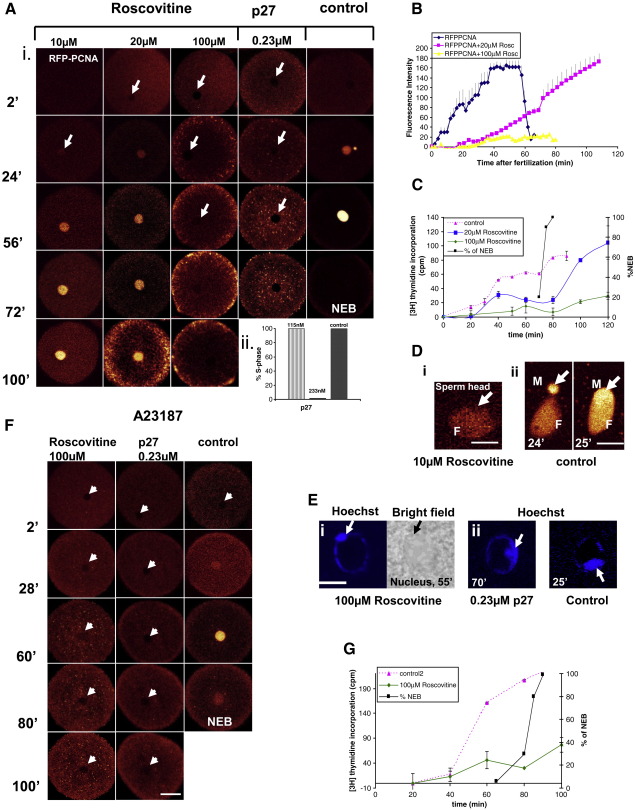
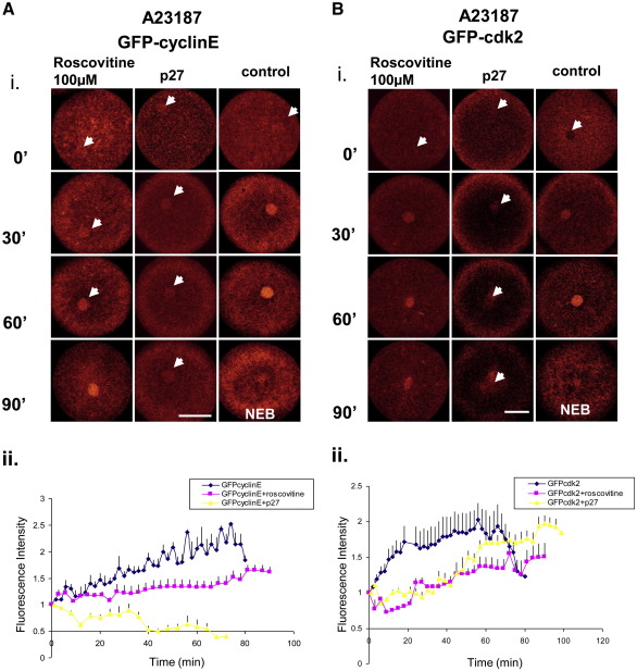
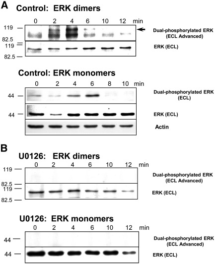
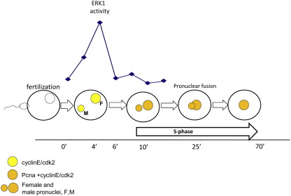
Sea urchins provide an excellent model for studying cell cycle control mechanisms governing DNA replication in vivo. Fertilization and cell cycle progression are tightly coordinated by Ca(2+) signals, but the mechanisms underlying the onset of DNA replication after fertilization remain less clear. In this study we demonstrate that calcium-dependent activation of ERK1 promotes accumulation of cyclinE/cdk2 into the male and female pronucleus and entry into first S-phase. We show that cdk2 activity rises quickly after fertilization to a maximum at 4 min, corresponding in timing to the early ERK1 activity peak. Abolishing MAP kinase activity after fertilization with MEK inhibitor, U0126, substantially reduces the early peak of cdk2 activity and prevents cyclinE and cdk2 accumulation in both sperm pronucleus and zygote nucleus in vivo. Both p27(kip1) and roscovitine, cdk2 inhibitors, prevented DNA replication suggesting cdk2 involvement in this process in sea urchin. Inhibition of cdk2 activity using p27(kip1) had no effect on the phosphorylation of MBP by ERK, but completely abolished phosphorylation of retinoblastoma protein, a cdk2 substrate, indicating that cdk2 activity is downstream of ERK1 activation. This pattern of regulation of DNA synthesis conforms to the pattern observed in mammalian somatic cells.


Figures

Fig. 1
Fig. 1
Nuclear localization of cdk2, cyclinE, and PCNA following fertilization or ionophore activation. (A) Eggs were injected with GFP-cdk2 and fertilized. GFP-cdk2 localises in the nucleus within first 3 min AF. (B, D) Eggs were co-injected with GFP-cyclinE or GFP-cdk2 together with RFP-PCNA. GFP-cdk2 and GFP-cyclinE co-localise with RFP-PCNA in fertilized eggs after 7–10 min. GFP-cyclinE and GFP-cdk2 can be detected however, in the female pronucleus within 3 min of insemination, well before pronuclear fusion. C,E. Quantified fluorescence intensity: (C) GFP-cyclinE and GFP-cdk2 co-localize with RFP-PCNA in the decondensing male pronucleus after 7–10 min; (E) parallel accumulation of GFP-cyclinE, GFP-cdk2 and RFP-PCNA in female and zygote nucleus. (F) Accumulation of GFP-cyclinE (i) and GFP-cdk2 (ii) in ionophore-activated eggs. Both proteins accumulate in the female nucleus after calcium elevation in the absence of sperm. Bar, 50 μm. Each graph represents mean and SEM of 4 individual experiments, except for GFP-cyclinE where 5 different experiments were made. For each experiment at least 3 different females were used.
Fig. 2
Fig. 2
Cdk2 activity during the first zygotic cell cycle depends on ERK1 activation. (A) Cdk2 activity, measured in whole-cell extracts using C-terminus of Rb protein as substrate, increased rapidly at 4 and 70 min AF. (B) p27kip1 inhibits the control cdk2 peak activities at 4, 12 and 70 min AF by 99, 90 and 96.6% respectively. (C and D) 100 μM of U0126 significantly reduced the first (C, at 4 min AF) and second peak (D, at 70 min AF) of cdk2 activity. (E) Control and U0126-inhibited ERK1 activity measured in the same cell extracts. The peak corresponds in time to cdk2 early peak in activity. (F) (i) The effect of p27kip1 on the phosphorylation of Rb and MBP in vitro. See text for details. (ii) p27kip1 inhibits Rb and MBP phosphorylation by 97% and 18% respectively. All results come from 3 independent experiments. Whole-cell extracts were obtained from 3 different females.
Fig. 3
Fig. 3
MAP kinase activation is essential for proper accumulation of cyclinE/cdk2 in male and female pronuclei, and the zygotic nucleus. (A) (i and ii) Eggs were injected with GFP-cyclinE (i) or GFP-cdk2 (ii) before fertilization. Embryos treated with MEK-specific inhibitor U0126, were unable to accumulate cyclinE or cdk2 in the zygotic nucleus when compared to control. Circles indicate the nucleus. Arrows indicate the position of centrosomes and chromosomes or nucleus when bright field was shown; bar 50 μm. The difference between images A (i) and A (ii) reflects the degree of averaging during laser scanning: in A (i) each scan was averaged times 8 and A (ii) each scan was averaged times 4. (B) (i and ii) Comparison between nuclear fluorescence intensity in control and U0126-treated embryos. (i) Embryos injected with GFP-cyclinE and (ii) GFP-cdk2. Each graph represents mean and SEM of 3 individual experiments for GFP-cdk2 and GFP-cdk2 with U1026, for GFP-cyclinE +/− U0126 5 different experiments were made. For each experiment at least 3 different females were used. (C) (i and ii) Inhibition of GFP-cyclinE (i) and GFP-cdk2 (ii) accumulation in the male pronucleus in U0126-treated embryos. Hoechst staining indicates the position of sperm chromatin after pronuclear fusion. Arrows in control eggs indicate the position of male pronucleus at pronucler fusion. Bar 10 μm.
Fig. 4
Fig. 4
Cdk2 activity is necessary for S-phase progression. (A) (i) RFP-PCNA was co-injected with 0.23 μM p27kip1 or alone before fertilization. Roscovitine (10, 20, 100 μM) was added 30 min before fertilization. DNA replication was abolished by both 0.23 μM p27kip1 and 100 μM roscovitine. Bar 50 μm. (ii) Concentration-dependent inhibition of DNA replication by p27kip1. S-phase continues with lower doses of p27 but is inhibited by 0.23 μM p27. 3 different experiments for each dose were used. B. Nuclear RFP-PCNA fluorescence intensity in control embryos and in the presence of 20 or 100 μM roscovitine. Data shown as mean and SEM of 4 individual experiments. (C) [3H]-Thymidine incorporation in control embryos and in the presence of 20 and 100 μM roscovitine. (D) (i) Inhibition of RFP-PCNA accumulation in the male pronucleus by 10 μM roscovitine. (ii) control images of RFP-PCNA during pronuclear fusion, Bar 10 μm, M—male, F—female pronucleus. (E) Abnormal pronuclear fusion after treatment with 100 μM roscovitine (i) and lack of sperm decondensation in embryos injected with 0.23 μM p27 (ii) compared to typical pronuclear fusion at 25 min. Bar, 10 μm. (F) Lack of DNA replication (showed by RFP-PCNA incorporation into nucleus) in roscovitine-treated or p27kip1-co-injected ionophore-activated eggs. (G) Thymidine incorporation after ionophore alone or together with roscovitine treatment. Data in C and G shown as mean and SEM of three individual experiments. Bar, 50 μm.
Fig. 5
Fig. 5
Cdk2 inhibitors affect accumulation of cyclinE and cdk2 in the female pronucleus of ionophore-activated eggs. (A and B) Representative images (i) and fluorescence intensity plots of nuclear fluorescence (ii) of GFP-cyclinE and GFP-cdk2 accumulation in the nucleus of ionophore-activated eggs after treatment with 100 μM roscovitine or injection of 0.23 μM p27. We also noted that when inhibitors were added or injected activation with ionophore took slightly longer to detect. This may be the reason why at time 0 (activation) we could detect very low fluorescence derived from GFP-cyclinE and GFP-cdk2. Roscovitine decreases the amount of cyclinE and cdk2 incorporated into nucleus. Nuclear accumulation of cyclinE when injected with p27 is almost completely abolished (Ai–ii). Cdk2 accumulation, however, while very low during first 40 min post-activation, increased rapidly after this time to about 70–80% of control (Bi–ii). Graphs represent mean and SEM of 4 individual experiments for cyclinE and n = 3 for cdk2 from three different females. Bar 50 μm.
Fig. 6
Fig. 6
(A, B) The formation of fully activated ERK bis-phosphodimers (arrow) corresponds exactly to the measured maximum in ERK activity. Phosphorylated ERK monomers do not contribute to the extremely high ERK activity measured at 2 and especially 4 min AF. (A) Control embryos. (B) U0126 inhibition at 15 min before fertilisation. The whole-cell extracts used for measurement of ERK1 and cdk2 activities were subjected to WB using anti-dual-phosphorylated ERK (upper panels) or anti-ERK antibodies (lower panels). Equal amounts of total protein were loaded on each gel (see actin loading control). Times AF and molecular markers are shown. Note that highly sensitive ECL was used for active ERK-dimer visualisation (arrow). Non-denaturing gel was used to maintain ERK as a dimer.
Fig. 7
Fig. 7
Proposed pathway controlling S-phase progression in sea urchin early embryos. Raf/MEK/ERK1 pathway controls the increase of cdk2 activity after fertilization and its translocation into male and female nucleus. At 6 min AF ERK1 activity drops down, DNA replication starts and S-phase progresses.

Similar articles

Cited by

References

    1. Bastians H., Townsley F.M., Ruderman J.V. The cyclin-dependent kinase inhibitor p27Kip1 induces N-terminal proteolytic cleavage of cyclin A. PNAS (US). 1998:15374–15381. - PMC - PubMed
    1. Blow J.J., Dutta A. Preventing re-replication of chromosomal DNA. Natl. Rev. Mol. Cell Biol. 2005;6:476–486. - PMC - PubMed
    1. Cameron L., Poccia D. In vitro development of the sea urchin male pronucleus. Dev. Biol. 1994;162:568–578. - PubMed
    1. Carroll D.J., Albay D.T., Hoang K.M., O'Neil F.J., Kumano M., Foltz K.R. The relationship between calcium, MAP kinase, and DNA synthesis in sea urchin egg at fertilization. Dev. Biol. 2000;217:179–191. - PubMed
    1. Chambard J.C., Lefloch R., Pouyssegur J., Lenormand P. ERK implication in cell cycle regulation. Bioch. Bioph. Acta November. 2006;17:1–12. - PubMed

Publication types

Substances