. 2009 Dec 1;164(2):877-85.
doi: 10.1016/j.neuroscience.2009.08.002. Epub 2009 Aug 7.

The supplementary motor area contributes to the timing of the anticipatory postural adjustment during step initiation in participants with and without Parkinson's disease

Affiliations

The supplementary motor area contributes to the timing of the anticipatory postural adjustment during step initiation in participants with and without Parkinson's disease

J V Jacobs et al. Neuroscience. .

Abstract

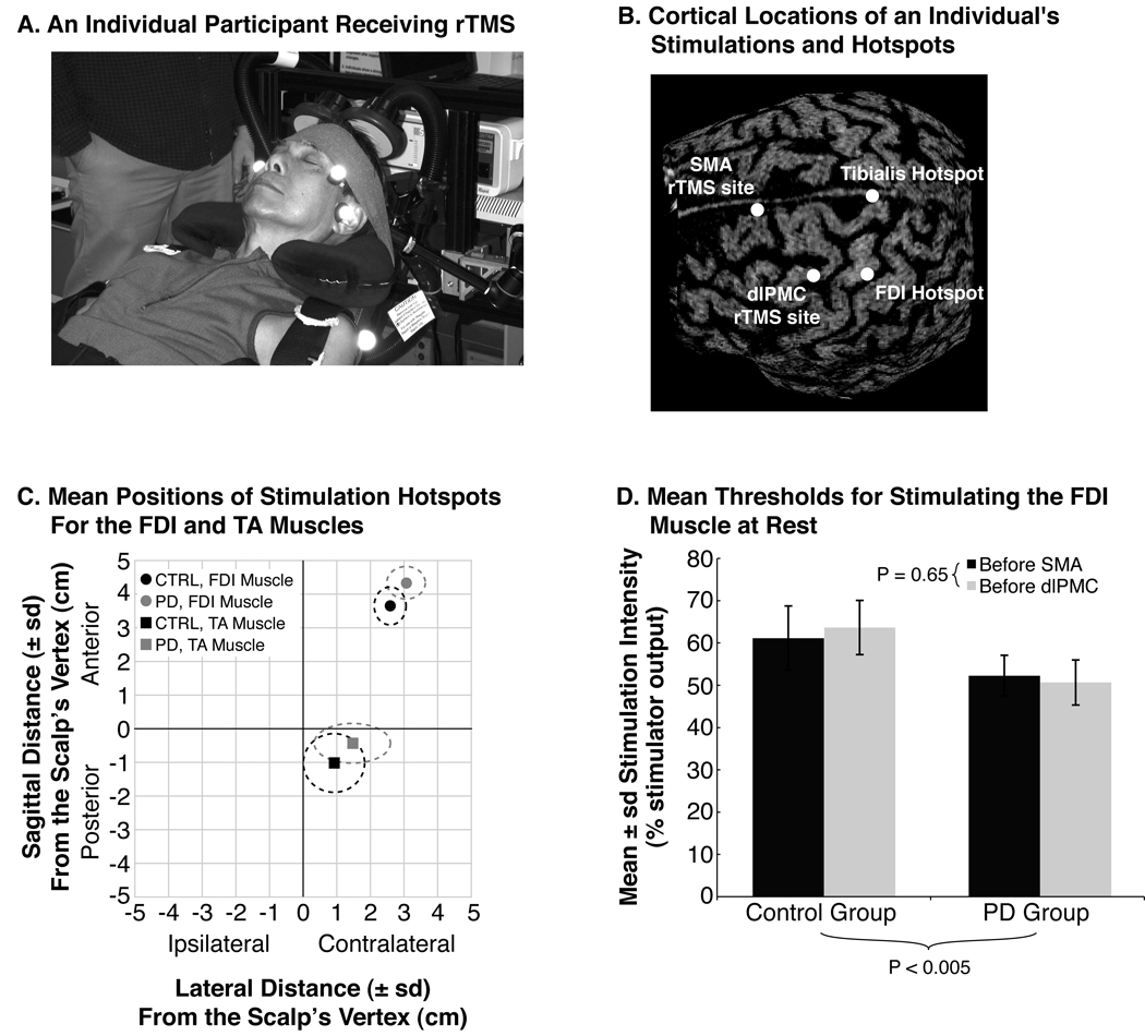
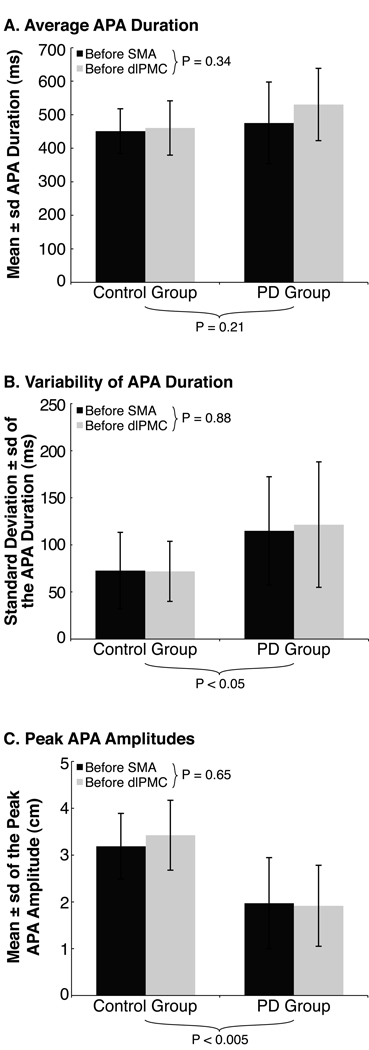
The supplementary motor area (SMA) is thought to contribute to the generation of anticipatory postural adjustments (APAs, which act to stabilize supporting body segments prior to movement), but its precise role remains unclear. In addition, participants with Parkinson's disease (PD) exhibit impaired function of the SMA as well as decreased amplitudes and altered timing of the APA during step initiation, but the contribution of the SMA to these impairments also remains unclear. To determine how the SMA contributes to generating the APA and to the impaired APAs of participants with PD, we examined the voluntary steps of eight participants with PD and eight participants without PD, before and after disrupting the SMA and dorsolateral premotor cortex (dlPMC), in separate sessions, with 1-Hz repetitive transcranial magnetic stimulation (rTMS). Both groups exhibited decreased durations of their APAs after rTMS over the SMA but not over the dlPMC. Peak amplitudes of the APAs were unaffected by rTMS to either site. The symptom severity of the participants with PD positively correlated with the extent that rTMS over the SMA affected the durations of their APAs. The results suggest that the SMA contributes to the timing of the APA and that participants with PD exhibit impaired timing of their APAs, in part, due to progressive dysfunction of circuits associated with the SMA.


Figures

Fig. 1
Fig. 1
Characteristics of rTMS. (A) A participant receiving rTMS over the SMA. The participant sat reclined in an adjustable dental chair with a memory foam pillow supporting his head and neck. An elastic band was also wrapped around the forehead to prevent excessive movement. The air-cooled coil of the Magstim rapid device was held in place by an adjustable clamp. (B) Image-guided TMS, demonstrating the cortical locations of muscle hotspots and of rTMS. (C) The average (SD) hotspot locations for the participants with PD (gray symbols and dashed lines) and the participants without PD (black symbols and dashed lines), relative to the vertex of the skull. The squares represent the hotspots for stimulating the TA muscle, and circles represent those for stimulating the FDI muscle. (D) The average (SD) rest motor thresholds of the FDI muscle during the sessions for rTMS over the SMA (dark gray bars) and dlPMC (light gray bars). Repetitive TMS was applied at 80 % of each participant’s rest motor threshold for that day’s session. The p-value below the chart represents the main effect of group differences, and the p-value next to the inset legend represents the main effect of session differences.
Fig. 2
Fig. 2
Characteristics of the APA prior to rTMS. The charts illustrate each group’s average (SD) (A) APA duration, (B) inter-trial variability of APA duration, and (C) peak APA amplitude prior to rTMS during the SMA (dark gray bars) and dlPMC (light gray bars) sessions. P-values below the charts represent main effects for group differences, those next to the inset legends represent main effects for session differences.
Fig. 3
Fig. 3
Effects of rTMS on the APA. (A) An example of shortened APA duration for one trial after rTMS over the SMA from an individual with PD. The horizontal axis represents time relative to APA onset, and the vertical axis represents the lateral displacement of the CoP for individual trials before stimulation (the thin gray curves), the average of trials before stimulation (the thin black curve), and for the first trial after SMA stimulation (the thick gray curve). Negative displacements are directed toward the participant’s swing limb. (B) Average APA durations by trial for all participants, demonstrating how APA durations decreased for only one trial after SMA stimulation; no group effects were evident. The black line with squares represents the mean APA durations from the session of rTMS over the SMA; the gray line with circles, the session of rTMS over the dlPMC. The asterisk highlights the first trial after rTMS because this trial was significantly different from others. (C) Average peak APA amplitudes by trial for all participants, demonstrating how APA amplitudes were smallest for the sessions’ first trials compared to subsequent trials (asterisk); no significant changes following rTMS and no group effects were evident.
Fig. 4
Fig. 4
A scatter plot illustrating a significant correlation among the PD participants’ disease severity (measured by lower-body motor UPDRS scores) and the extent that rTMS over the SMA affected APA durations. The circles represent the values for individual participants with PD; the Xs, those of the participants without PD. Although the UPDRS was not assessed for those without PD, their values are depicted with an assumed UPDRS score of zero. The horizontal axis has been changed so that positive values represent a decrease in APA duration following rTMS in order to illustrate a positive correlation among disease severity and the effect of rTMS on APA duration.

References

    1. Bestmann S, Baudewig J, Siebner HR, Rothwell JC, Frahm J. BOLD MRI responses to repetitive TMS over human dorsal premotor cortex. Neuroimage. 2005;28:22–29. - PubMed
    1. Bloem BR, Grimbergen YA, Cramer M, Willemsen M, Zwinderman AH. Prospective assessment of falls in Parkinson’s disease. J Neurol. 2001;248:950–958. - PubMed
    1. Bloem BR, Hausdorff JM, Visser JE, Giladi N. Falls and freezing of gait in Parkinson’s disease: a review of two interconnected, episodic phenomena. Mov Disord. 2004;19:871–884. - PubMed
    1. Boylan LS, Pullman SL, Lisanby SH, Spicknall KE, Sackeim HA. Repetitive transcranial magnetic stimulation to SMA worsens complex movements in Parkinson’s disease. Clin Neurophysiol. 2001;112:259–264. - PubMed
    1. Braak H, Del Tredici K, Bratzke H, Hamm-Clement J, Sandmann-Keil D, Rüb U. Staging of the intracerebral inclusion body pathology associated with idiopathic Parkinson’s disease (preclinical and clinical stages) J Neurol. 2002;249 Suppl 3:1–5. - PubMed

Publication types