. 2009 Aug 7;138(3):449-62.
doi: 10.1016/j.cell.2009.05.035.

The ectopic expression of Pax4 in the mouse pancreas converts progenitor cells into alpha and subsequently beta cells

Affiliations

The ectopic expression of Pax4 in the mouse pancreas converts progenitor cells into alpha and subsequently beta cells

Patrick Collombat et al. Cell. .

Abstract

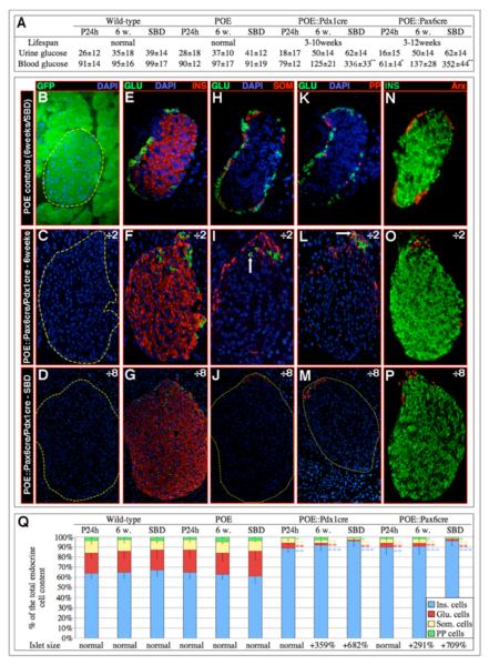
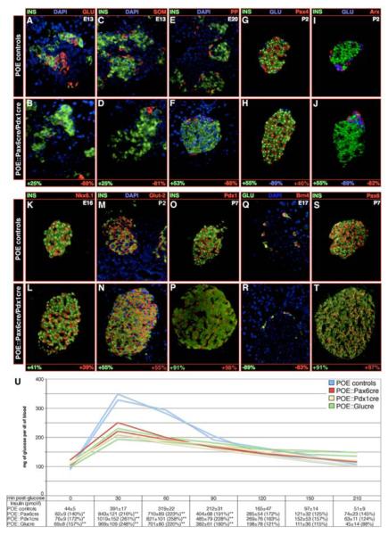
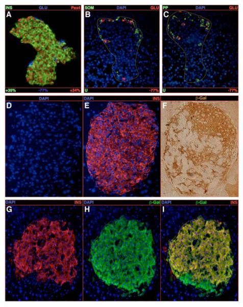
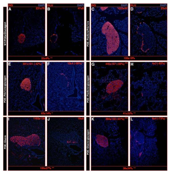
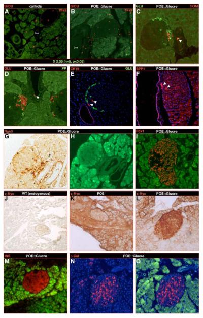
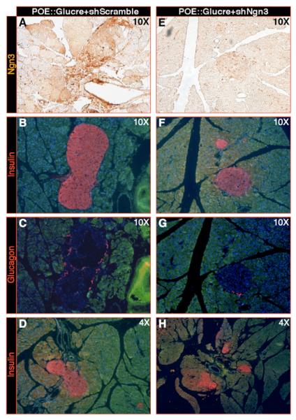
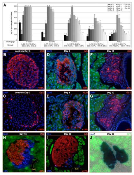
We have previously reported that the loss of Arx and/or Pax4 gene activity leads to a shift in the fate of the different endocrine cell subtypes in the mouse pancreas, without affecting the total endocrine cell numbers. Here, we conditionally and ectopically express Pax4 using different cell-specific promoters and demonstrate that Pax4 forces endocrine precursor cells, as well as mature alpha cells, to adopt a beta cell destiny. This results in a glucagon deficiency that provokes a compensatory and continuous glucagon+ cell neogenesis requiring the re-expression of the proendocrine gene Ngn3. However, the newly formed alpha cells fail to correct the hypoglucagonemia since they subsequently acquire a beta cell phenotype upon Pax4 ectopic expression. Notably, this cycle of neogenesis and redifferentiation caused by ectopic expression of Pax4 in alpha cells is capable of restoring a functional beta cell mass and curing diabetes in animals that have been chemically depleted of beta cells.


Figures

Figure 1
Figure 1. The overexpression of Pax4 in Pdx1 or Pax6 expression domains promotes the genesis of oversized islets mainly containing insulin-expressing cells
(A) Characterization of the lifespan and glycemia (in mg/dl) of POE::Pdx1cre and POE::Pax6cre mice 1 day and 6 weeks postpartum, as well as short before death (SBD). Note that initially, these animals are hypo- and subsequently hyperglycemic. (B-P) Immunohistochemical analysis of the islets of double-transgenic mice. A dramatic increase in islet size and insulin-expressing cell mass is evident 6 weeks postpartum (C, F, I, L, O) and is even more pronounced SBD (D, G, J, M, P) as compared to representative control islets (B, E, H, K, N). Concurrently, a loss of α- (E-M), δ- (H-J), PP- (K-M), and Arx-labeled (N-P) cells is evident, but, interestingly, few of these remain detectable at one pole of the islet, some co-expressing the glucagon and somatostatin (arrow in I) or PP (arrow in L) hormones. (Q) A quantification of the endocrine cell alterations ascertains these observations (also see Table S2). For the purpose of clarity, the magnification of double-transgenic islets is twice (C, F, I, L, O) or eight times (D, G, J, M, P) reduced compared to controls. (n>11, * P<0.05, ** P<0.01, *** P<0.001, all values expressed as means ± standard error of the mean).
Figure 2
Figure 2. Insulin-expressing cells detected in POE::Pdx1cre and POE::Pax6cre pancreata exhibit a β-cell identity
(A-T) Characterization of POE::Pdx1cre and POE::Pax6cre hormone-expressing cells. Sections of double-transgenic pancreata (of the indicated ages) were stained using the mentioned antibodies, the number of labeled cells counted and reported to the count obtained in POE control animals. The values are expressed in percentage of change in labeled cell number compared to controls. All values are statistically significant with P values lower than or equal to 0.05. Note that the numbers of insulin-expressing cells are dramatically increased in double-transgenic animals at all examined stages (A-P, S-T) and this, as early as E13 (A-B), compared to controls. Concurrently, the mean contents of α- (A-B, G-J, Q-R), δ- (C-D), PP- (E-F) and Arx- (I-J) marked cells appear drastically reduced. A thorough analysis of endocrine cell-specific markers demonstrates that, all insulin-producing cells clearly express Pax4 (G-H), as well as the β-cell markers Nkx6.1 (K-L), Glut-2 (M-N) and Pdx1 (O-P). These are negative for the α-cell specific factor Brn-4 (Q-R), whereas, all endocrine cells express Pax6 (S-T). (U) An improved glucose tolerance and insulin release are highlighted in 3-week-old double-transgenic mice as compared to controls POE mice (n>3, ***P<0.001, **P<0.01, * P<0.05). Each picture is representative of at least 4 independent animals; note that, for the purpose of clarity, a POE::Pax6cre islet is displayed in H.
Figure 3
Figure 3. Conversion of glucagon-expressing cells into insulin-producing cells upon Pax4 ectopic expression
(A-C) Quantification of the endocrine cell content alterations upon ectopic expression of the Pax4 gene in glucagon-producing cells in 1-week-old animals. A clear increase in the number of insulin-/Pax4-labeled cells at the expense of glucagon-expressing cells is highlighted (A), whereas the δ- and PP-cell contents are found unchanged (B-C). Note the accumulation of the remaining glucagon-marked cells at one pole of the islet. (D-F) The detection of insulin- and β-galactosidase-expressing cells on serial sections demonstrates that numerous insulin-labeled cells do express the β-galactosidase gene that normally marks glucagon-positive cells. (G-I) The same observation is made in 6-week old animals using co-immunofluorescence. All reported values are statistically significant (P<0.05, n=5). See Table S2 for a detailed analysis.
Figure 4
Figure 4. Exogenous glucagon supplementation prevents islet overgrowth
Six-week old pancreata of the indicated genotypes were sectioned and stained using anti-insulin (A, C, E, G, I, K) or glucagon (B, D, F, H, J, L) antibodies. The values at the top right corner of each photograph represent the number of hormone-producing cells per square centimeter of pancreas. The counts reported underneath each picture set represent variations in islet surface (estimated in silico) using POE::Pax6cre/Pdx1cre pancreata (C-D) as reference for photographs A-H and POE::Glucre pancreata (I-J) as reference for photographs I-L. Note the overall diminution in islet size and glucagon+ cell content, as well as the drastic decrease in insulin-expressing cell number (E-H compared to C-D and K-L compared to I-J) in animals supplemented with glucagon for 3 weeks compared to untreated ones (*P<0.05, **P<0.01, n=3).
Figure 5
Figure 5. Progenitor cells may be induced and converted into glucagon and subsequently into insulin-expressing cells
Next to a pulse of BrdU at three weeks of age and examination a week later, a quantitative analysis established a 2.35 increase in the number of proliferating cells in POE::Glucre pancreas compared to POE controls (A-B). Importantly, most BrdU-labeled cells are not detected within the islets, but rather near or within the duct epithelium (B). It is worth noticing that this location corresponds to the cluster of glucagon-producing cells consistently observed in this genotype. Within the duct epithelium, scattered endocrine cells could be detected, some co-expressing the glucagon and sometimes somatostatin hormones (arrowheads in C-D). Along the same line, islet cells positive for the duct marker genes CK19 and SPP1 are observed adjacent to the duct epithelium (arrowheads in E-F). Notably, the duct lining also appears to contain numerous cells positive for the proendocrine marker gene Ngn3 (G-H), but negative for Pdx1 (I). (J-L) Infection of controls (K) and double-transgenic (L-O) pancreata using a construct containing a CMV promoter driving the constitutive expression of a c-Myc tag (the latter being not expressed in wild-type tissues - J). Due to the method used, two weeks post-infection, most exocrine cells are labeled by the virus, whereas only very few islet cells are (islet underlined in K). Importantly, a majority of double-transgenic endocrine cells appear marked by the virus, suggesting their ductal or acinar origin (L). These cells express the insulin hormone (M) and are β-galactosidase-positive (N-O), indicating that they once expressed the glucagon hormone. (H-I, M, O: Green fluorescence corresponding to residual GFP expression after bleaching of the sections).
Figure 6
Figure 6. Knockdown of Ngn3 prevents the Pax4-mediated β-cell hyperplasia
(A-H) Infection of POE::Glucre animals using lentiviruses producing either an shRNA targeting Ngn3 transcripts (E-H - Xu et al., 2008) or producing a scrambled shRNA (based on the former; A-D). Two weeks post-infection, Ngn3 knockdown pancreata (at the indicated magnifications) display an efficient 61% diminution in Ngn3 production (Xu et al., 2008, A, E and Table S6), but also a 69% decrease in insulin- (B, F, D, H) or glucagon- (C, G) expressing cell numbers (Table S6) compared to scramble-infected counterparts.
Figure 7
Figure 7. Pax4 ectopic expression promotes the reconstitution of the insulin-expressing cell mass upon β-cell depletion
(A) Following streptozotocin injection at the indicated age (starting age), the glycemia and survival of the treated animals was followed for two months. Note that animals older than four weeks of age become diabetic and die as a consequence of the obliteration of β-cells, the same being true for all controls (data not shown). Importantly, in younger animals, a steady recovery leading to normoglycemia is observed next to a peak in glucose levels (all values are expressed as means ± standard error of the mean). (B-J) Islet cell contents in 4-week-old POE::Glucre streptozotocin- (D-J) or sham- (B-C) treated animals were quantified (see at the bottom of the concerned pictures) 3 days (B-E), 10 days (F-G), 16 days (H) and 60 days (I-J) days post-injection. Three days post-injection, the β-cell mass present in controls (B-C) is almost entirely lost in streptozotocin-treated mice (D-E), the only insulin labeling being observed in areas devoid of nuclei, suggesting a detection of hormone released from killed β-cells. A 15% increase in glucagon-producing cells is highlighted 10 days post-injection compared to controls, most of these cells neighboring duct structures (G). Importantly, few insulin-labeled cells are detected (F). This trend is ascertained at day 16 (H) with a major increase in the insulin-expressing cell number. A co-detection with the glucagon hormone does not indicate any co-expression. Finally, at day 60, the β-cell mass appears statistically normal as compared to control animals (I compared to B-C), most of the cells present in the islet expressing the β-galactosidase enzyme marking glucagon-producing cells (J).

Comment in

Similar articles

Cited by

References

    1. Ahlgren U, Jonsson J, Edlund H. The morphogenesis of the pancreatic mesenchyme is uncoupled from that of the pancreatic epithelium in IPF1/PDX1-deficient mice. Development. 1996;122:1409–1416. - PubMed
    1. Ahlgren U, Jonsson J, Jonsson L, Simu K, Edlund H. beta-cell-specific inactivation of the mouse Ipf1/Pdx1 gene results in loss of the beta-cell phenotype and maturity onset diabetes. Genes Dev. 1998;12:1763–1768. - PMC - PubMed
    1. Ashery-Padan R, Zhou X, Marquardt T, Herrera P, Toube L, Berry A, Gruss P. Conditional inactivation of Pax6 in the pancreas causes early onset of diabetes. Dev Biol. 2004;269:479–488. - PubMed
    1. Blume N, Skouv J, Larsson LI, Holst JJ, Madsen OD. Potent inhibitory effects of transplantable rat glucagonomas and insulinomas on the respective endogenous islet cells are associated with pancreatic apoptosis. J Clin Invest. 1995;96:2227–2235. - PMC - PubMed
    1. Brun T, Franklin I, St-Onge L, Biason-Lauber A, Schoenle EJ, Wollheim CB, Gauthier BR. The diabetes-linked transcription factor PAX4 promotes {beta}-cell proliferation and survival in rat and human islets. J Cell Biol. 2004;167:1123–1135. - PMC - PubMed

Publication types

MeSH terms