. 2009 Aug 7;138(3):463-75.
doi: 10.1016/j.cell.2009.05.026.

Telomere extension occurs at most chromosome ends and is uncoupled from fill-in in human cancer cells

Affiliations

Telomere extension occurs at most chromosome ends and is uncoupled from fill-in in human cancer cells

Yong Zhao et al. Cell. .

Abstract

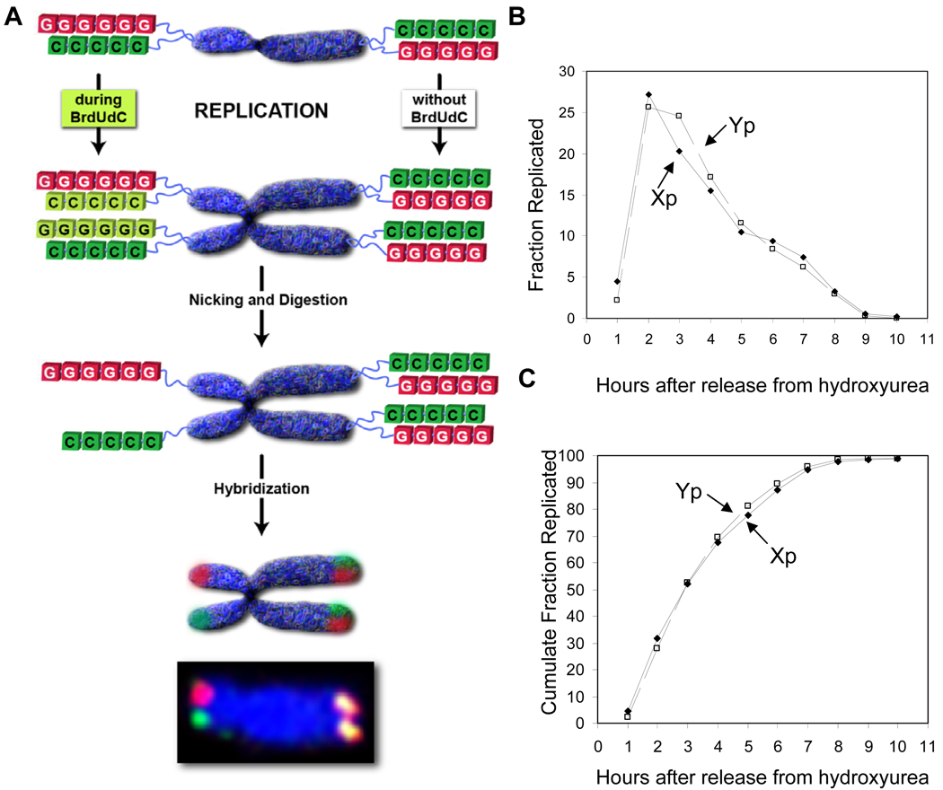
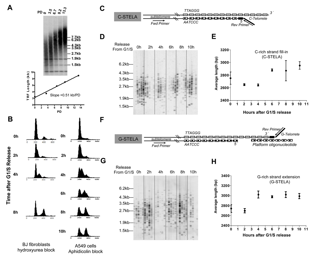
Telomeres are thought to be maintained by the preferential recruitment of telomerase to the shortest telomeres. The extension of the G-rich telomeric strand by telomerase is also believed to be coordinated with the complementary synthesis of the C strand by the conventional replication machinery. However, we show that under telomere length-maintenance conditions in cancer cells, human telomerase extends most chromosome ends during each S phase and is not preferentially recruited to the shortest telomeres. Telomerase rapidly extends the G-rich strand following telomere replication but fill-in of the C strand is delayed into late S phase. This late C-strand fill-in is not executed by conventional Okazaki fragment synthesis but by a mechanism using a series of small incremental steps. These findings highlight differences between telomerase actions during steady state versus nonequilibrium conditions and reveal steps in the human telomere maintenance pathway that may provide additional targets for the development of anti-telomerase therapeutics.


Figures

Figure 1
Figure 1. Timing of replication of the Xp/Yp telomere
(A) Schema for the determination of replication timing using ReDFISH. Only one of two strands survives nicking/digestion of hemisubstituted DNA. The fraction (B) or cumulative total (C) of replicating Xp/Yp telomeres was quantitated from metaphase spreads after hourly pulses. 70% of Xp/Yp telomeres replicate during hours 2–4 after the release of hydroxyurea synchronized BJ fibroblasts.
Figure 2
Figure 2
Dissociation of telomerase extension and C-strand fill-in in human cancer cells (A) Telomere restriction fragments (TRF) analysis of hTERT retrovirus infected A549 cells. Telomere length increased at ~500bp/PD. (B) FACS analysis of DNA content in BJ fibroblasts and A549 cells synchronized at G1/S and every 2h after release into S until G2 (10h after release). (C) Schematic of C-STELA for C-rich strand length of the Xp/Yp telomeres. Reverse PCR-priming sequences terminating in short 7-nucleotide permutations of the hexameric C-rich sequence are annealed to the G-rich overhang, ligated, then amplified using a unique primer in the Xp/Yp subtelomere and the artificial reverse primer. (D) Five sample lanes representing five different C-STELA amplifications of the Xp/Yp telomere in A549 cells from each time point after G1/S release. (E) Quantitation of the data in (D). The average size of ~100 telomeres was determined for each time point. +/− SD of 3 independent experiments. (F) Schematic of the G-STELA for G-rich strands length. Annealing of a C-rich "platform" oligonucleotides permits alignment and ligation of the G-telorette sequences (G) Five sample lanes at each time point after G1/S release of G-STELA amplifications of the Xp/Yp telomere. (E) Quantitation of the data in (G). The average size of ~100 telomeres was determined for each time point. +/− SD of 3 independent experiments.
Figure 3
Figure 3. Overhang elongation during S phase
(A). DSN analysis on agarose gels of the hTERT overexpressing A549 cells used in Fig. 2 at different times following release from a G1/S block. Overhang size increased by several hundred nt during S phase before returning to the size in unsynchronized cells at 10 hours after release. (B) DNA collected following the release of synchronized Hela cells into S-phase was digested with DSN before and after digestion with Exo1 to remove 3' overhangs and analyzed on polyacrylamide gels. The weight-averaged mean overhang size increased during S until returning to their G1 size at S/G2. (C) The results of (B) were confirmed by in-gel hybridization to native ds telomeres. The gel was then denatured to determine the total telomere signal, and the values expressed as a ratio between the native and denatured signals. The results are plotted as a % of the maximum value. +/− SD for three experiments. (D) Hela DNA replicating during a 30 minute BrdU pulse 3 hours after release or during 8 hours of continuous label following release from G1/S were separated on CsCl gradients. Leading and lagging telomeric daughters were resolved under both conditions. (E) DSN overhang analysis of the leading and lagging fractions isolated from (D). The weight-averaged mean size from two independent experiments is shown below the gel. (G) Quantitation of the data in (E) is shown as a cumulative fraction, so that the distribution of sizes can be distinguished from the average. A cumulative fraction value of 0.7 at a specific size means that 70% of the overhangs are shorter and 30% are longer than that specific size. The length of both leading and lagging overhangs increased within 30 minutes of replication compared to at 8 hours when significant fill-in had begun in this experiment (see B).
Figure 4
Figure 4. Direct detection of telomerase addition during S phase
(A) Strategy to study telomerase extension on lagging daughter telomeres. The leading strand daughter is thought to be initially blunt ended, but is shown following processing to generate a small G-rich overhang. Leading daughter overhangs are always fully BrdU substituted regardless of telomerase action. Prior to C-strand fill-in, lagging overhangs extended by telomerase would contain a mixture of thymidine and BrdU substituted segments. (B) CsCl gradient resolution of telomeric overhangs with and without BrdU incorporation. DNA from synchronized Hela cells before and after release into BrdU containing medium for 48h was digested with DSN and the overhangs were analyzed on CsCl gradients. (C) G1/S synchronized Hela cells were released into S phase for 3h in the presence of BrdU. Genomic DNA was DSN digested then spun on CsCl gradients. An intermediate density peak is seen. Synchronized normal telomerase negative fibroblasts (BJ) released into S phase for 4h in the presence of BrdU did not show an intermediate peak. Hela cells treated with the telomerase inhibitor GRN163L for 7 days before synchronization and release in BrdU also lacked an intermediate density peak (D) Aliquots of the CsCl gradient from the first panel of (C) were slot blotted follow by hybridizing with either C-rich (left) or G-rich (right) telomeric probes. Samples were collected from the bottom of the gradient, and loaded in two columns on each slot blot. The positions of the thymidine and BrdU peaks are labeled. Signals above background were only seen with the C-rich probe for the G-rich overhang sequence.
Figure 5
Figure 5
Telomerase extension versus C-Strand fill-in in Hela cells. (A) FACS analysis of synchronized Hela cells released into S phase for different times in the presence of BrdU. (B) Time course of overhang density distribution during S phase. The intermediate peak only becomes heavier late in S phase. (C) Pulse-chase strategy for the early replicating cohort of telomeres. (D) Time course of the overhang density distribution of pulse-chased early replicating telomeres.
Figure 6
Figure 6
Fraction of lagging daughter telomeres extended by telomerase in one cell cycle. (A) DNA from G1/S synchronized Hela cells released in BrdU for 4 hours was separated on CsCl gradients. (B) Four fractions from the lagging strand peak in (A) were pooled and digested with DSN. The resulting overhangs were then analyzed on a second CsCl gradient. The thymidine peak represents those replicated ends that were not extended by telomerase. (C) Deconvolution of the peaks in (B) assuming a Gaussian distribution for each overhang class. (D) Replotting of the data in (C) after normalization for the estimated size of the extended vs non-extended overhangs (see text). ~70% of Hela telomeres are extended each cell cycle. (E) Telomeres from synchronized H1299 cells released for 4 hours in BrdU were separated into unreplicated, lagging and leading fractions. (F) 100% of lagging overhangs exhibited intermediate densities indicating extension by telomerase. (G) Overhangs from unreplicated telomeres showed no evidence of BrdU incorporation. (H) Overhangs from leading strand daughter telomeres were fully substituted with BrdU.
Figure 7
Figure 7
Model for the replication of mammalian telomeres. See text for details.

Comment in

References

    1. Adams AK, Holm C. Specific DNA replication mutations affect telomere length in Saccharomyces cerevisiae. Mol Cell Biol. 1996;16:4614–4620. - PMC - PubMed
    1. Adams Martin A, Dionne I, Wellinger RJ, Holm C. The function of DNA polymerase alpha at telomeric G tails is important for telomere homeostasis. Mol Cell Biol. 2000;20:786–796. - PMC - PubMed
    1. Bailey SM, Goodwin EH, Cornforth MN. Strand-specific fluorescence in situ hybridization: the CO-FISH family. Cytogenet Genome Res. 2004;107:14–17. - PubMed
    1. Baird DM, Rowson J, Wynford-Thomas D, Kipling D. Extensive allelic variation and ultrashort telomeres in senescent human cells. Nat Genet. 2003;33:203–207. - PubMed
    1. Bianchi A, Shore D. How telomerase reaches its end: mechanism of telomerase regulation by the telomeric complex. Mol Cell. 2008;31:153–165. - PubMed

Publication types

LinkOut - more resources