. 2009 Oct 30;393(3):717-34.
doi: 10.1016/j.jmb.2009.08.001. Epub 2009 Aug 8.

A method for helical RNA global structure determination in solution using small-angle x-ray scattering and NMR measurements

Affiliations

A method for helical RNA global structure determination in solution using small-angle x-ray scattering and NMR measurements

Jinbu Wang et al. J Mol Biol. .

Abstract

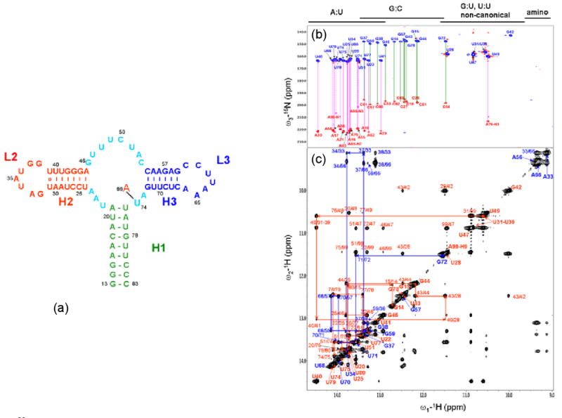
We report a "top-down" method that uses mainly duplexes' global orientations and overall molecular dimension and shape restraints, which were extracted from experimental NMR and small-angle X-ray scattering data, respectively, to determine global architectures of RNA molecules consisting of mostly A-form-like duplexes. The method is implemented in the G2G (from global measurement to global structure) toolkit of programs. We demonstrate the efficiency and accuracy of the method by determining the global structure of a 71-nt RNA using experimental data. The backbone root-mean-square deviation of the ensemble of the calculated global structures relative to the X-ray crystal structure is 3.0+/-0.3 A using the experimental data and is only 2.5+/-0.2 A for the three duplexes that were orientation restrained during the calculation. The global structure simplifies interpretation of multidimensional nuclear Overhauser spectra for high-resolution structure determination. The potential general application of the method for RNA structure determination is discussed.


Figures

Figure 1
Figure 1
The definition of duplex orientation (Φ,Θ) and phase ρ0 (a & b), simulated RDC-structural periodicity correlation curves (c) that are color-coded the same as in (a) and the definition of duplex axis (d and e). The shapes of the RDC-structural periodicity correlation curves (RDC waves) depend on the orientation and phase of duplexes. The shapes of the RDC-structural periodicity correlation curves depend on orientation (Θ,Φ) and the phase ρ0. The orientations and phases of duplexes colored in red, blue and cyan are (Θ,Φ,ρ0), are (45°, 45°, 0°), (60°, 300°, 120°) and (75°, 128°, 232°), respectively. The RDC waves are simulated using Da=-15.6 Hz and R=0.22. The A-form duplexes were generated using Discovery Studio 2.0 (Accelrys Software, Inc.). (d) Each ball represents a phosphate atom (P) and a basepair PP vector stands for the vector from 3′ P to 5′ P in the same base pair. Big bold black arrow shows the duplex orientation. Big bold black arrow indicates the duplex orientation. (e) The vector ΔPi¯=pi+1pj+1¯pipj¯ belongs to the plane perpendicular to the duplex axis. The cross product ni = ΔPi ⊗ ΔPi+1 is the normal of the plane perpendicular to the duplex axis, which is parallel to the duplex axis. The duplex orientation is calculated as an average of the normals.
Figure 1
Figure 1
The definition of duplex orientation (Φ,Θ) and phase ρ0 (a & b), simulated RDC-structural periodicity correlation curves (c) that are color-coded the same as in (a) and the definition of duplex axis (d and e). The shapes of the RDC-structural periodicity correlation curves (RDC waves) depend on the orientation and phase of duplexes. The shapes of the RDC-structural periodicity correlation curves depend on orientation (Θ,Φ) and the phase ρ0. The orientations and phases of duplexes colored in red, blue and cyan are (Θ,Φ,ρ0), are (45°, 45°, 0°), (60°, 300°, 120°) and (75°, 128°, 232°), respectively. The RDC waves are simulated using Da=-15.6 Hz and R=0.22. The A-form duplexes were generated using Discovery Studio 2.0 (Accelrys Software, Inc.). (d) Each ball represents a phosphate atom (P) and a basepair PP vector stands for the vector from 3′ P to 5′ P in the same base pair. Big bold black arrow shows the duplex orientation. Big bold black arrow indicates the duplex orientation. (e) The vector ΔPi¯=pi+1pj+1¯pipj¯ belongs to the plane perpendicular to the duplex axis. The cross product ni = ΔPi ⊗ ΔPi+1 is the normal of the plane perpendicular to the duplex axis, which is parallel to the duplex axis. The duplex orientation is calculated as an average of the normals.
Figure 2
Figure 2
Schematic drawing of the secondary structure of the riboA RNA (a) and its 2D NMR spectra, (b) HNN-COSY spectrum of 15N-labeled riboA sample, and (c) 2D NOESY spectrum of non-labeled riboA in the imino and partial amino proton region. In (a), the ligand adenine is labeled as A99 in the diagram and is in a close contact with U74. In (b), AU pairs are linked with magenta lines, and GC with green lines. Dashed lines denote missing acceptor because the ligand adenine is the acceptor but not isotope-labeled. In (c), two NOE-walking paths are shown in red and blue.
Figure 3
Figure 3
Imino 15N-1H IPAP spectra of the 15N-labeled riboA, recorded in an anisotropic solution containing about 0.5 mM RNA and 9.7 mg/ml pf1 alignment medium. The spectra were recorded at 25 °C on a VARIAN Inova spectrometer operating at proton frequency 800 MHz and equipped with a cryogenic triple resonance probe with a z-gradient. The imino peaks are generally well resolved.
Figure 4
Figure 4
The dipolar waves for H1, H2 and H3 in riboA from the simultaneous fits. Four DROs simultaneously satisfy each wave. The solid curves are back-calculated waves using experimental Da, R, orientation (Θ,Φ) and phase ρ0 from simultaneous fits using the program ORIENT (see the text and SI) and structural parameters of a standard A-form duplex. The experimental RDCs are drawn as circles. The deviation of experimental RDCs from the curves at several residues may be due in part to deviation from the ideal A-form geometry. RDC of residue G78 was not used due to large error caused by peak overlapping.
Figure 5
Figure 5
Combinations of possible orientations for duplexes H1 and H2 (a-d), and duplexes H1 and H3 (e-h) for riboA. Duplex H1, H2 and H3 are shown in green, red and in blue, and linkers in cyan. In general, two conformations in each combination are not viable because steric clashes between a linker and duplexes, as in conformations b, d, f and g. The coordinates of the possible conformers were generated by using the programs BLOCK and PACK and dawn using Pymol (DeLano, W.L. DeLano Scientific, San Carlos, CA, USA. http://www.pymol.org).
Figure 6
Figure 6
The shape-assisted identification of the correct combination of DRO. The comparisons of the riboA molecular envelope and the initial structures built from viable orientation combinations leads to the conclusion that either a parallel or an anti-parallel arrangement of the three duplexes, conformer ae, can possibly fit into the envelope.
Figure 7
Figure 7
(a) A two dimensional topological drawing (left) and the three dimensional starting structure (right) that was generated using the G2G toolkit. The orientations and phases (Θ,Φ,ρ0) of the duplexes are given alongside the duplexes. The broken lines represent linker residues. Close contacts between H2 and H3 involving imino protons were unambiguously detected in the NOESY spectrum (NMR experiments section). The three duplexes have correct orientations and phases which determine the terminal positions of linkers and loops. The initial relative positions of the duplexes were arranged to be approximately consistent with the dimensions of the envelope. The hairpin loops and linkers, derived from the MOSAIC library, were added with arbitrary conformations so long as they can be connected to the ends of the three duplexes via 3′O to a phosphate atom (SI) without a sharp twist. The deviations from covalent geometry, such as abnormal bond lengths and angles at the joint junctions, were corrected in the regularization process in the SA calculation. (b) The initial fold of the riboA structure that was calculated using the rigid-body SA refinement protocol. The backbone RMSDs between the top 10% initial fold structures and the X-ray structure are 2.5 ± 0.2 and 3.4 ± 0.3 Å for the three duplexes and the whole molecule, respectively.
Figure 7
Figure 7
(a) A two dimensional topological drawing (left) and the three dimensional starting structure (right) that was generated using the G2G toolkit. The orientations and phases (Θ,Φ,ρ0) of the duplexes are given alongside the duplexes. The broken lines represent linker residues. Close contacts between H2 and H3 involving imino protons were unambiguously detected in the NOESY spectrum (NMR experiments section). The three duplexes have correct orientations and phases which determine the terminal positions of linkers and loops. The initial relative positions of the duplexes were arranged to be approximately consistent with the dimensions of the envelope. The hairpin loops and linkers, derived from the MOSAIC library, were added with arbitrary conformations so long as they can be connected to the ends of the three duplexes via 3′O to a phosphate atom (SI) without a sharp twist. The deviations from covalent geometry, such as abnormal bond lengths and angles at the joint junctions, were corrected in the regularization process in the SA calculation. (b) The initial fold of the riboA structure that was calculated using the rigid-body SA refinement protocol. The backbone RMSDs between the top 10% initial fold structures and the X-ray structure are 2.5 ± 0.2 and 3.4 ± 0.3 Å for the three duplexes and the whole molecule, respectively.
Figure 8
Figure 8
The ensemble of global structures of the riboA RNA determined using the G2G “top-down” method, a SAXS curve comparison and the adenine ligand position. (a) The front (center) and side (left and right) views of the superimposed riboA backbone structures (top 10% lowest energy) superimposed within the envelope. The X-ray crystal structure is in cyan, the G2G structures in red, and the average structure of the ensemble in blue. The local structures of the hairpin loops and linker between H2 and H3 are relatively poorly defined. The backbone RMSDs of the ensemble (top 10% of 100 structures calculated) relative to the mean is about 0.8 Å. (b) The superimposed correlation plots between the imino RDCs that were back-calculated from the ensemble of the top 10% lowest energy structures using the Powell-Grid-search method and the experimental RDCs. (c) The correlation plot between the imino RDCs that were back-calculated from the regularized average structure of the ensemble of the top 10% and the experimental RDCs. (d) The correlation plot between the imino RDCs that were back-calculated from the X-ray crystal structure (1Y26) and the experimental RDCs. (e) The comparison of experimental (circle), back-calculated SAXS curves based on the ensemble (red), average (blue) and the X-ray crystal structure (cyan) and the RMSD between the first and the third is about 0.29 ± 0.04, which is calculated based on the logarithms of the normalized (i.e., I(q=0)=1.0) SAXS intensities. (f) The comparison of experimental PDDF (black), and those for the ensemble (red), the average (blue) and the X-ray crystal structure (cyan). (g) The comparison of the adenine ligand positions in the X-ray crystal structure (cyan) and the average structure of the top 10% lowest energy G2G structures. The adenine ligands are shown in sphere mode. The adenine position was defined by five close contacts that were detected in NOESY spectra (Figures 2c and S4 in SI).
Figure 8
Figure 8
The ensemble of global structures of the riboA RNA determined using the G2G “top-down” method, a SAXS curve comparison and the adenine ligand position. (a) The front (center) and side (left and right) views of the superimposed riboA backbone structures (top 10% lowest energy) superimposed within the envelope. The X-ray crystal structure is in cyan, the G2G structures in red, and the average structure of the ensemble in blue. The local structures of the hairpin loops and linker between H2 and H3 are relatively poorly defined. The backbone RMSDs of the ensemble (top 10% of 100 structures calculated) relative to the mean is about 0.8 Å. (b) The superimposed correlation plots between the imino RDCs that were back-calculated from the ensemble of the top 10% lowest energy structures using the Powell-Grid-search method and the experimental RDCs. (c) The correlation plot between the imino RDCs that were back-calculated from the regularized average structure of the ensemble of the top 10% and the experimental RDCs. (d) The correlation plot between the imino RDCs that were back-calculated from the X-ray crystal structure (1Y26) and the experimental RDCs. (e) The comparison of experimental (circle), back-calculated SAXS curves based on the ensemble (red), average (blue) and the X-ray crystal structure (cyan) and the RMSD between the first and the third is about 0.29 ± 0.04, which is calculated based on the logarithms of the normalized (i.e., I(q=0)=1.0) SAXS intensities. (f) The comparison of experimental PDDF (black), and those for the ensemble (red), the average (blue) and the X-ray crystal structure (cyan). (g) The comparison of the adenine ligand positions in the X-ray crystal structure (cyan) and the average structure of the top 10% lowest energy G2G structures. The adenine ligands are shown in sphere mode. The adenine position was defined by five close contacts that were detected in NOESY spectra (Figures 2c and S4 in SI).

References

    1. Fire A, Xu S, Montgomery MK, Kostas SA, Driver SE, Mello CC. Potent and specific genetic interference by double-stranded RNA in Caenorhabditis elegans. Nature. 1998;391:806–811. - PubMed
    1. Tucker BJ, Breaker RR. Riboswitches as versatile gene control elements. Curr Opin Struct Biol. 2005;15:342–348. - PubMed
    1. Kruger K, Grabowski PJ, Zaug AJ, Sands J, Gottschling DE, Cech TR. Self-splicing RNA: autoexcision and autocyclization of the ribosomal RNA intervening sequence of Tetrahymena. Cell. 1982;31:147–157. - PubMed
    1. Blackburn EH. Telomerases. Annu Rev Biochem. 1992;61:113–129. - PubMed
    1. Moore PB, Steitz TA. The involvement of RNA in ribosome function. Nature. 2002;418:229–235. - PubMed

Publication types