. 2009 Oct 30;184(1):104-9.
doi: 10.1016/j.jneumeth.2009.07.031. Epub 2009 Aug 8.

Chronic electrical stimulation of cultured hippocampal networks increases spontaneous spike rates

Affiliations

Chronic electrical stimulation of cultured hippocampal networks increases spontaneous spike rates

Gregory J Brewer et al. J Neurosci Methods. .

Abstract

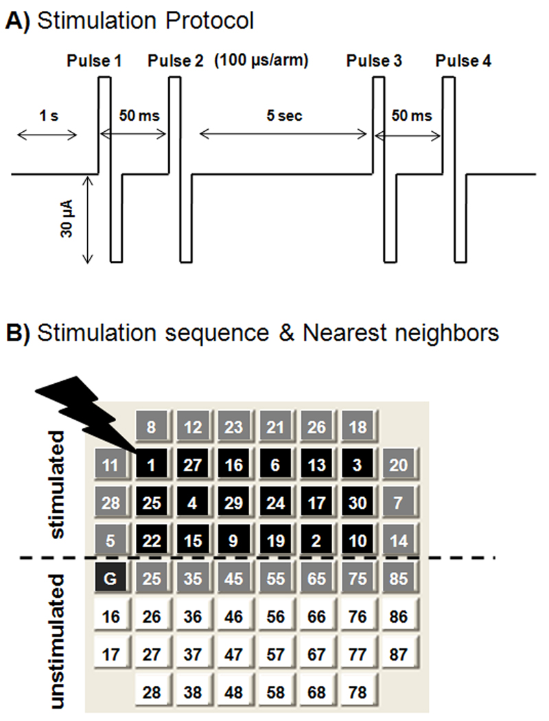
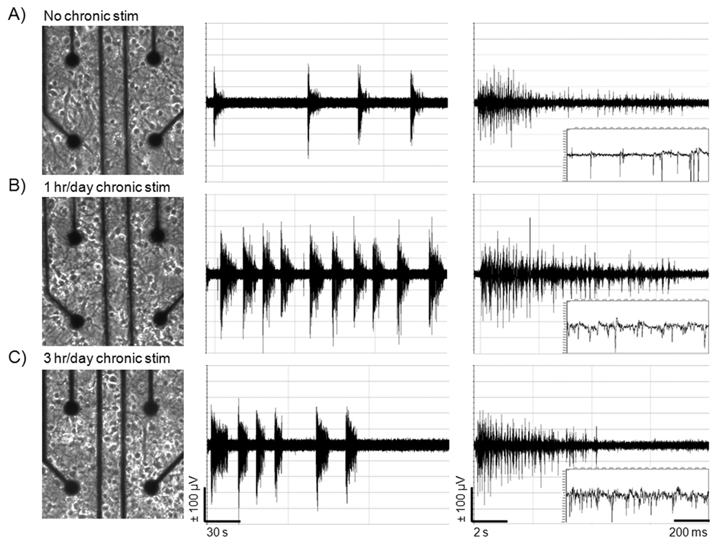
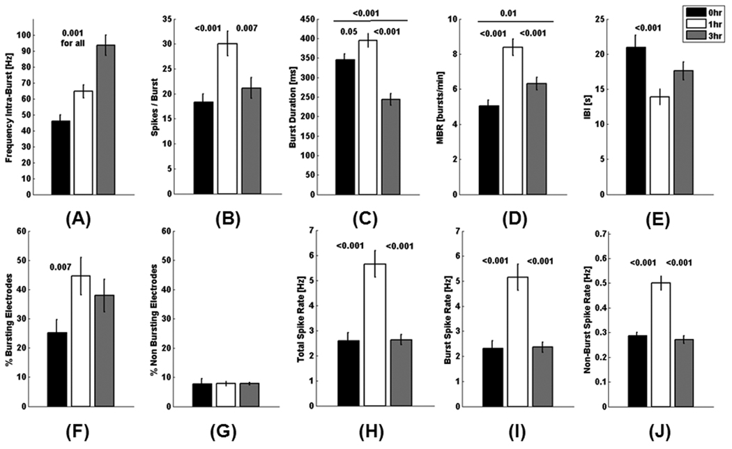
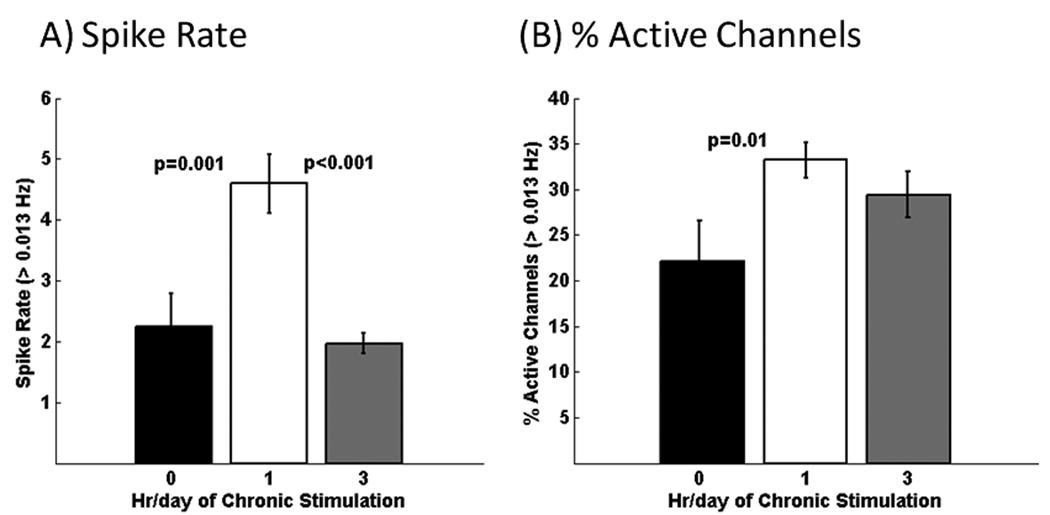
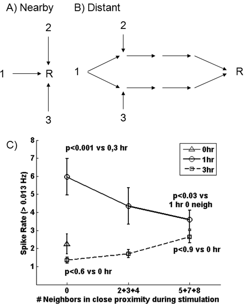
We chronically stimulated hippocampal networks in culture for either 0, 1 or 3h/day between 7 and 22 days in culture in an effort to increase spontaneous spike rates and to give these networks some portion of external stimuli that brain networks receive during their formation. Chronic electrical stimulation of hippocampal networks on multi-electrode arrays (MEAs) increased spike rates 2-fold after 3 weeks of culture compared to cultures that received no external stimulation prior to recording. More than 90% of the spikes for all experimental conditions occurred within bursts. The frequency of spikes within a burst increased with time of stimulation during culture up to 2-fold higher (90Hz) compared to networks without chronic stimulation. However, spontaneous overall spike rates did not correlate well with the amount of stimulation either as h/day or proximity to the limited number of stimulation sites due to shorter burst duration with 3h/day stimulation. The results suggest that chronic stimulation applied during network development recruits activity at 50% more electrodes and enables higher rates of spontaneous activity within bursts in cultured hippocampal networks.


Figures

Fig. 1
Fig. 1
Chronic stimulation protocol applied to determine effect of external stimuli on spike rates during time in culture. A) A train of two paired pulses, 50 ms between single pulses in a pair, 5 sec apart was delivered to a single electrode, followed by switching to a second electrode1 sec later. B) The switching was repeated to each of 30 electrodes in top half of array. This process was repeated for either 1 or 3 hr/day for a total of either 2100 or 6300 pulses per array/day over 8 days. The program switches channels in a pseudorandom sequence after each train to avoid stimulation of any two adjacent electrodes consecutively and to minimize short-term plasticity. Black squares represent electrodes with 5, 7, or 8 neighbours in close proximity during stimulation. Gray squares represent electrodes with 2, 3, or 4 neighbours in close proximity and white squares represent electrodes with zero neighbours in close proximity.
Fig. 2
Fig. 2
Chronic stimulation increases total bursts and burst frequency without morphological changes in neurons on MEAs compared to unstimulated conditions. A) Neuron distribution on an MEA after 21 days of culture in NbActiv4 for unstimulated condition, B) 1 hr/day chronic stimulation, and C) 3 hr/day chronic stimulation. Corresponding amplifier outputs are shown to the right. Frequency of bursts is highest with 1 hr/day chronic stimulation. Frequency of spikes within a burst is highest at 3 hr/day stimulation.
Fig. 3
Fig. 3
Chronic stimulation during culture increases burst spike frequency, spikes/burst, burst duration and total bursts compared to unstimulated condition after 3 weeks. Data represents analysis of channels with one or more bursts. The average response for each electrode from the active electrodes from 5 arrays/condition. p values represent student’s t-test (n=144, 254, and 216 active electrodes from 5 arrays/condition of 0, 1, and 3 hr/day stimulation). A) Spike frequency within a burst increases with duration of chronic stimulation. B) Spikes/ burst are highest with 1 hr/day chronic stimulation, but similar to control at 3 hr/day chronic stimulation. C) Burst duration is longest with 1 hr/day chronic stimulation and lowest with 3 hr/day chronic stimulation. D) 1 hr/day chronic stimulation produces a 2-fold increase in mean burst rate (bursts/ minute) above the unstimulated condition. E) Interburst interval (IBI) of 21 seconds for the unstimulated condition is 1-1/2 fold higher than the IBI of cultures chronically stimulated for 1 hr/day. F) Percentage of bursting electrodes nearly doubles with 1hr/day chronic stimulation and increases 50% with 3 hr/day chronic stimulation compared to unstimulated condition. G) Electrodes containing non-bursting spikes amounted to less than 10 percent of the total active electrodes for all conditions. H, I) Total spike rates and burst spike rates were more than 2-fold higher with 1 hr/day chronic stimulation compared to unstimulated and 3 hr/day stimulated conditions. J) Non-bursting spike rates were nearly 2-fold higher with 1 hr/day chronic stimulation compared to unstimulated and 3 hr/day stimulated conditions.
Fig. 4
Fig. 4
Considering all channels with >0.013 Hz activity, both bursting plus non-bursting, A) chronic stimulation for 1 hr/day produces 2-fold higher overall spike rates. B) Either chronic stimulation condition caused a 30–50% higher percentage of active electrodes per MEA compared to the unstimulated condition (n=221, 332, and 294 active electrodes from 5 arrays/condition of 0, 1, and 3 hr/day stimulation).
Fig. 5
Fig. 5
Impact of stimulation proximity on spike rate. A) Recording sites at close proximity to simulating sites 1, 2, or 3 condition these sites for higher spontaneous spike rates. Alternatively, B) pathways that get recruited by chronic stimulation more often converge at a distant electrode to increase spike rates. C) Measures of electrodes on MEA cultures receiving 1 hr/day (circle) of chronic stimulation produce 3-fold higher spike rates for recording sites distant in proximity to the stimulating electrode (0 nearby stimuli), compared to unstimulated or 3 hr/day chronically stimulated cultures. Spike rates decrease for electrodes with more frequent adjacent stimulation. For 3 hr/day stimulation (squares), the spike rate was similar to all sites on arrays without chronic stimulation (triangle), but tended to increase for sites that received close-proximity stimuli.

References

    1. Boehler MD, Wheeler BC, Brewer GJ. Added astroglia promote greater synapse density and higher activity in neuronal networks. Neuron Glia Biol. 2007;3:127–140. - PMC - PubMed
    1. Brewer GJ, Boehler MD, Jones TT, Wheeler BC. NbActiv4 medium improvement to Neurobasal/B27 increases neuron synapse densities and network spike rates on multielectrode arrays. J. Neurosci. Meth. 2008a;170:181–187. - PMC - PubMed
    1. Brewer GJ, Boehler MD, Pearson RA, Demaris AA, Ide AN, Wheeler BC. Neuron network activity scales exponentially with synapse density. J. Neural Eng. 2009;6:14001. - PMC - PubMed
    1. Chang JC, Brewer GJ, Wheeler BC. Neuronal network structuring induces greater neuronal activity through enhanced astroglial development. J. Neural Eng. 2006;3:217–226. - PubMed
    1. Chiappalone M, Novellino A, Vajda I, Vato A, Martinoia S, van Pelt J. Burst detection algorithms for the analysis of spatio-temporal patterns in cortical networks of neurons. Neurocomputing. 2005;65–66:653–662.

Publication types

LinkOut - more resources