. 2009 Oct 9;284(41):28128-28136.
doi: 10.1074/jbc.M109.041483. Epub 2009 Aug 7.

Critical role for tetrahydrobiopterin recycling by dihydrofolate reductase in regulation of endothelial nitric-oxide synthase coupling: relative importance of the de novo biopterin synthesis versus salvage pathways

Affiliations

Critical role for tetrahydrobiopterin recycling by dihydrofolate reductase in regulation of endothelial nitric-oxide synthase coupling: relative importance of the de novo biopterin synthesis versus salvage pathways

Mark J Crabtree et al. J Biol Chem. .

Abstract

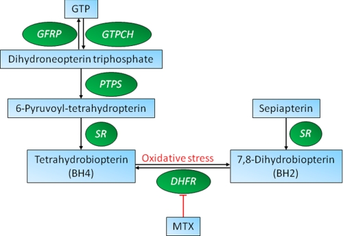
Tetrahyrobiopterin (BH4) is a required cofactor for the synthesis of nitric oxide by endothelial nitric-oxide synthase (eNOS), and BH4 bioavailability within the endothelium is a critical factor in regulating the balance between NO and superoxide production by eNOS (eNOS coupling). BH4 levels are determined by the activity of GTP cyclohydrolase I (GTPCH), the rate-limiting enzyme in de novo BH4 biosynthesis. However, BH4 levels may also be influenced by oxidation, forming 7,8-dihydrobiopterin (BH2), which promotes eNOS uncoupling. Conversely, dihydrofolate reductase (DHFR) can regenerate BH4 from BH2, but the functional importance of DHFR in maintaining eNOS coupling remains unclear. We investigated the role of DHFR in regulating BH4 versus BH2 levels in endothelial cells and in cell lines expressing eNOS combined with tet-regulated GTPCH expression in order to compare the effects of low or high levels of de novo BH4 biosynthesis. Pharmacological inhibition of DHFR activity by methotrexate or genetic knockdown of DHFR protein by RNA interference reduced intracellular BH4 and increased BH2 levels resulting in enzymatic uncoupling of eNOS, as indicated by increased eNOS-dependent superoxide but reduced NO production. In contrast to the decreased BH4:BH2 ratio induced by DHFR knockdown, GTPCH knockdown greatly reduced total biopterin levels but with no change in BH4:BH2 ratio. In cells expressing eNOS with low biopterin levels, DHFR inhibition or knockdown further diminished the BH4:BH2 ratio and exacerbated eNOS uncoupling. Taken together, these data reveal a key role for DHFR in eNOS coupling by maintaining the BH4:BH2 ratio, particularly in conditions of low total biopterin availability.


Figures

FIGURE 1.
FIGURE 1.
Schematic representation of the BH4 recycling pathway and eNOS coupling. BH4 is synthesized from GTP via a series of reactions involving GTPCH, 6-pyruvoyl-tetrahydropterin synthase, sepiapterin reductase (SR) and DHFR. DHFR activity can be inhibited by MTX. GFRP, GTP cyclohydrolase feedback regulatory protein. PTPS, 6-pyruvoyl-tetrahydropterin synthase.
FIGURE 2.
FIGURE 2.
DHFR overexpression in sEnd.1 endothelial cells. A, DHFR levels are increased by 5-fold after transfection of human DHFR cDNA in sEnd.1 endothelial cells as shown by Western blotting using a DHFR-specific antibody, as detailed under “Experimental Procedures.” Representative blots are shown; n = 3. DHFR overexpression did not significantly affect intracellular BH4 levels (B), total biopterin levels (C), the production of nitric oxide as demonstrated by A23187-induced conversion of arginine to citrulline (D), or the amount of eNOS-derived superoxide (E); n = 3 or 6 for each experiment.
FIGURE 3.
FIGURE 3.
Inhibition of endogenous DHFR activity by MTX:biopterin levels. sEnd.1 cells were exposed to MTX (1 μm) for 16 h at 37 °C, and intracellular biopterin levels were quantified by HPLC as detailed under “Experimental Procedures.” A, exposure to MTX did not affect intracellular eNOS, DHFR, or GTPCH (GCH) protein levels. B, exposure of sEnd.1 cells to MTX significantly decreased BH4 levels (*, p < 0.05), whereas a marked accumulation of BH2 (**, p < 0.001) (C) and biopterin (**, p < 0.001) (D) was observed. E, no significant change in total biopterin was demonstrated. F, BH4:BH2 ratio was significantly decreased after treatment of sEnd.1 cells with MTX (**, p < 0.001, n = 3 for all experiments).
FIGURE 4.
FIGURE 4.
Inhibition of endogenous DHFR activity by MTX:nitric oxide and superoxide production. A23187-induced conversion of arginine to citrulline was used as a measure of eNOS activity. A, eNOS activity (as indicated by the inhibition of citrulline accumulation by N-methyl arginine; NMA) was attenuated in the presence of MTX in sEnd.1 cells (*, p < 0.05). B, after exposure to MTX (1 μm) for 16 h, cells were exposed to dihydroethidium (25 μm) for 20 min, and accumulation of 2-hydroxyethidium was measured by HPLC with fluorescence detection. MTX exposure markedly elevated cellular superoxide production (**, p < 0.01), which was significantly inhibited after pretreatment of cells with the NOS inhibitor, l-NAME (C) (100 μm; *, p < 0.05). n = 6 for each experiment.
FIGURE 5.
FIGURE 5.
Genetic knockdown of DHFR and GTPCH protein by siRNA. sEnd.1 cells were transfected with DHFR- and GCH-specific, control GAPDH, or scrambled nonspecific siRNA as described under “Experimental Procedures.” A, DHFR and GTPCH-specific siRNAs alone or in combination knocked down protein levels by more than 90%. Importantly, GTPCH and GAPDH knockdown as well as scrambled control siRNAs did not have any effect on DHFR protein levels. B, Intracellular BH4 levels were decreased 40 and 95% by DHFR- and GTPCH-specific siRNAs, respectively (*, p < 0.05). C, a 2-fold increase in BH2 levels was induced by DHFR-specific siRNA (*, p < 0.05), and a 95% reduction was observed after exposure of sEnd.1 cells to GTPCH-specific siRNA (**, p < 0.001). Combined knockdown of GTPCH and DHFR resulted in a 4-fold increase in BH2 when compared with cells exposed to GTPCH-specific siRNA alone (†, p < 0.05). D, total biopterins were unchanged by DHFR-specific siRNA and reduced by ∼95% after exposure of sEnd.1 endothelial cells to GTPCH-specific siRNA or when combined with DHFR-specific siRNA (**, p < 0.001). E, intracellular BH4:BH2 ratio was markedly decreased by DHFR-, but not GTPCH-specific siRNA exposure (*, p < 0.05). Importantly, the BH4:BH2 ratio was further decreased when endothelial cells were exposed to both GTPCH- and DHFR-specific siRNAs combined (†, p < 0.05). F, A23187-induced conversion of arginine to citrulline cells was diminished by DHFR (*, p < 0.05) and GTPCH-specific siRNAs (**, p < 0.001). G and H, eNOS-dependent superoxide production was induced by GTPCH (*, p < 0.05)- and DHFR (*, p < 0.05)-specific siRNA and when combined, led to a significantly increased production versus either GTPCH or DHFR siRNAs alone (**, p < 0.001). Blots shown are representative of three separate experiments. All biopterin and arginine to citrulline measurements were unaffected by GAPDH knockdown or scrambled control siRNAs (n = 6).
FIGURE 6.
FIGURE 6.
MTX exposure allows oxidation of BH4 in GCH cells. To test whether the effect of MTX had a differential effect at different amounts of GTPCH, a dose-response curve with doxycycline was carried out in GCH cells expressing tetracycline-regulatable GTPCH, and BH4 was quantified by HPLC. GCH cells demonstrated a dose-dependent “switch-off” of BH4 synthesis after exposure to increasing concentrations of doxycycline (DOX) for 5 days. Cells exposed to MTX (1 μm) for 16 h (open symbols, dashed line) exhibit significantly lower amounts of BH4 compared with control cells (*, p < 0.05) (A) and a concomitant increase in BH2 levels (*, p < 0.05) (B). n = 3 for each experiment.
FIGURE 7.
FIGURE 7.
DHFR regulates eNOS coupling in GCH/eNOS cells. GCH and GCH/eNOS cells in the presence and absence of doxycycline were exposed to MTX (1 μm) for 16 h at 37 °C, and intracellular biopterin levels were quantified by HPLC as detailed under “Experimental Procedures.” A, Western blotting shows relative abundance of eNOS, GTPCH, and DHFR in respective cell lysates. Importantly, eNOS is only observed in GCH/eNOS cells and GTPCH protein expression is abolished following exposure to doxycycline. B, BH4 levels were significantly reduced in GCH/eNOS cells after exposure to MTX (*, p < 0.05). C, inhibition of DHFR with MTX resulted in a marked elevation in BH2 levels compared with untreated control cells in both GCH and GCH/eNOS cells (*, p < 0.05). BH2 levels were also increased in GCH/eNOS cells treated with both doxycycline and MTX compared doxycycline alone (†, p < 0.05). D, in GCH cells MTX, but not doxycycline, exposure alone was sufficient to induce a striking decrease in BH4:BH2 ratio (#, p < 0.01). In GCH/eNOS cells, doxycycline decreased the BH4:BH2 ratio (*, p < 0.05), which was further exacerbated in the presence of MTX (§, p < 0.05, n = 6).
FIGURE 8.
FIGURE 8.
Inhibition of DHFR in GCH versus GCH/eNOS cells. After exposure to MTX (1 μm) for 16 h, cells were exposed to dihydroethidium (25 μm) for 20 min, and accumulation of 2-hydroxyethidium was measured by HPLC with fluorescence detection. A and C, MTX exposure markedly elevated cellular superoxide production (*, p < 0.05); however, this was not inhibited after pretreatment of cells with the NOS inhibitor, l-NAME (100 μm). B and D, MTX exposure markedly elevated cellular superoxide production (*, p < 0.05), which was significantly inhibited by l-NAME (100 μm) (†, p < 0.05, n = 6).

References

    1. Vásquez-Vivar J., Kalyanaraman B., Martásek P., Hogg N., Masters B. S., Karoui H., Tordo P., Pritchard K. A., Jr. (1998) Proc. Natl. Acad. Sci. U.S.A. 95, 9220–9225 - PMC - PubMed
    1. Chalupsky K., Cai H. (2005) Proc. Natl. Acad. Sci. U.S.A. 102, 9056–9061 - PMC - PubMed
    1. Hurshman A. R., Krebs C., Edmondson D. E., Huynh B. H., Marletta M. A. (1999) Biochemistry 38, 15689–15696 - PubMed
    1. Schmidt P. P., Lange R., Gorren A. C., Werner E. R., Mayer B., Andersson K. K. (2001) J. Biol. Inorg. Chem. 6, 151–158 - PubMed
    1. Vásquez-Vivar J., Martásek P., Whitsett J., Joseph J., Kalyanaraman B. (2002) Biochem. J. 362, 733–739 - PMC - PubMed

Publication types

MeSH terms