. 2009 Oct 2;284(40):27596-608.
doi: 10.1074/jbc.M109.047563. Epub 2009 Aug 7.

NEMO-binding domains of both IKKalpha and IKKbeta regulate IkappaB kinase complex assembly and classical NF-kappaB activation

Affiliations

NEMO-binding domains of both IKKalpha and IKKbeta regulate IkappaB kinase complex assembly and classical NF-kappaB activation

Laura A Solt et al. J Biol Chem. .

Abstract

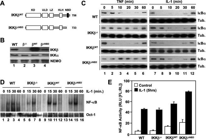
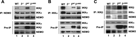
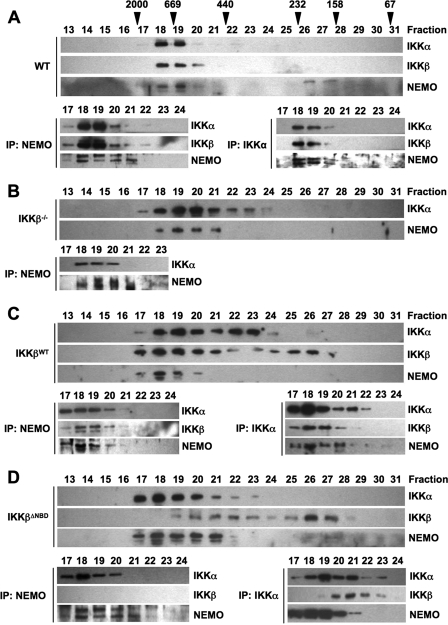
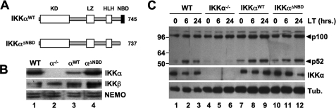
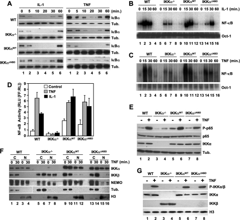
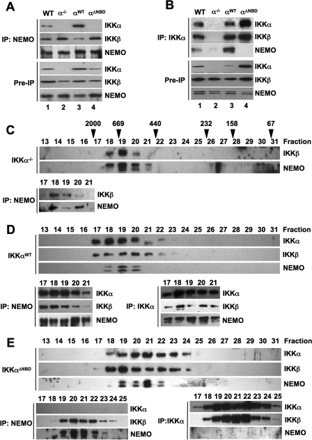
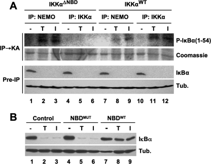
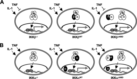
Proinflammatory NF-kappaB activation requires the IkappaB (inhibitor of NF-kappaB) kinase (IKK) complex that contains two catalytic subunits named IKKalpha and IKKbeta and a regulatory subunit named NF-kappaB essential modulator (NEMO). NEMO and IKKbeta are essential for tumor necrosis factor (TNF)-induced NF-kappaB activation, and we recently demonstrated that NEMO and IKKalpha are sufficient for interleukin (IL)-1-induced signaling. IKKalpha and IKKbeta both contain a functional NEMO-binding domain (NBD); however, the role of NEMO association with each kinase in NF-kappaB signaling and IKK complex formation remains unclear. To address this question, we stably reconstituted IKKalpha(-/-) and IKKbeta(-/-) murine embryonic fibroblasts (MEFs) with wild-type (WT) or NBD-deficient (DeltaNBD) versions of IKKalpha and IKKbeta, respectively. TNF-induced classical NF-kappaB activation in IKKbeta(-/-) MEFs was rescued by IKKbeta(WT) but not IKKbeta(DeltaNBD), whereas neither IKKbeta(WT) nor IKKbeta(DeltaNBD) affected IL-1-induced NF-kappaB signaling. As previously described, classical NF-kappaB transcriptional activity was absent in IKKalpha(-/-) cells. Reconstitution with either IKKalpha(WT) or IKKalpha(DeltaNBD) rescued both IL-1 and TNF-induced transcription, demonstrating that NEMO association is not required for IKKalpha-dependent regulation of NF-kappaB-dependent transcription. Stably expressed IKKalpha(WT) or IKKbeta(WT) associated with endogenous IKKs and NEMO in IKKalpha(-/-) or IKKbeta(-/-) MEFs, respectively, resulting in formation of the heterotrimeric IKKalpha-IKKbeta-NEMO complex. In contrast, although the IKKalpha(DeltaNBD) and IKKbeta(DeltaNBD) mutants associated with endogenous IKKs containing an NBD, these dimeric endogenous IKK-IKK(DeltaNBD) complexes did not associate with NEMO. These findings therefore demonstrate that formation of the heterotrimeric IKKalpha-IKKbeta-NEMO holocomplex absolutely requires two intact NEMO-binding domains.


Figures

FIGURE 1.
FIGURE 1.
The IKKβ NBD is required for TNF- but not IL-1-induced classical NF-κB activation. A, the structural domains of wild-type IKKβ (IKKβWT) and IKKβΔNBD are shown. KD, kinase domain; LZ, leucine zipper; ULD, ubiquitin-like domain; HLH, helix-loop-helix. IKKβΔNBD is a truncation mutant encompassing residues 1–733 that lacks the C-terminal 23 amino acids containing the NBD (13, 14). B, lysates from WT, IKKβ−/−−/−), IKKβWTWT), and IKKβΔNBDΔNBD) MEFs were immunoblotted using the antibodies indicated (right). C, WT, IKKβ−/−, IKKβWT, and IKKβΔNBD MEFs were incubated with either TNF (10 ng/ml) (left) or IL-1α (10 ng/ml) (right) for the times indicated, and then lysates were immunoblotted using anti-IκBα or anti-tubulin (Tub.) as a loading control. D, the same panel of MEFs was treated with IL-1α for the indicated times, and then nuclear extracts were prepared for EMSA. Assays were performed using either a consensus NF-κB binding site probe (top) or an Oct-1 probe as a loading control (bottom). E, MEFs were transiently transfected with the NF-κB-dependent reporter pBIIx-firefly luciferase together with β-actin Renilla luciferase. Twenty-four hours later, cells were either left untreated or treated for a further 5 h with IL-1α, and then NF-κB activity was determined by a dual luciferase assay.
FIGURE 2.
FIGURE 2.
IKKβΔNBD associates with endogenous IKKα but not with endogenous NEMO. IKK complexes in whole cell lysates of WT, IKKβ−/−−/−), IKKβWTWT), and IKKβΔNBDΔNBD) MEFs were immunoprecipitated (IP) using anti-NEMO (A), anti-IKKα (B), or anti-IKKβ (C). Immunoprecipitated material was immunoblotted using anti-IKKα, anti-IKKβ, and anti-NEMO as indicated (right). Samples of lysates saved prior to immunoprecipitation (Pre-IP) were immunoblotted using anti-IKKα, anti-IKKβ, and anti-NEMO as shown.
FIGURE 3.
FIGURE 3.
IKKβΔNBD is not incorporated into the tripartite IKK complex. S100 extracts from WT (A), IKKβ−/− (B), IKKβWT (C), and IKKβΔNBD MEFs (D) were fractionated by size exclusion chromatography. Fractions were immunoblotted using the antibodies indicated (left). The column was precalibrated, and the molecular weights of standard proteins are indicated above the appropriate fractions in A. Fractions containing the high molecular weight IKK complex were immunoprecipitated (IP) using either anti-NEMO or anti-IKKα. The resulting immunoblots from these immunoprecipitations are displayed below the fractionation profile for each cell type in A–D.
FIGURE 4.
FIGURE 4.
The IKKα NBD is not required for noncanonical NF-κB activation. A, the structural domains of wild-type IKKα (IKKαWT) and IKKαΔNBD are shown. KD, kinase domain; LZ, leucine zipper; HLH, helix-loop-helix. IKKαΔNBD is a truncation mutant encompassing residues 1–737 that lacks the C-terminal 8 amino acids containing the NBD (13, 14). B, lysates from WT, IKKα−/−−/−), IKKαWTWT), and IKKαΔNBDΔNBD) MEFs were immunoblotted using the antibodies indicated (right). C, WT, IKKα−/−, IKKαWT, and IKKαΔNBD MEFs were either untreated or incubated with LTα1β2 (LT) for the times indicated, and then lysates were immunoblotted using either anti-p100/p52 (top), anti-IKKα, or anti-tubulin (Tub.) as indicated (right).
FIGURE 5.
FIGURE 5.
IKKαΔNBD restores NF-κB-dependent transcriptional activity. A, WT, IKKα−/−, IKKαWT, and IKKαΔNBD MEFs were treated with either IL-1α (10 ng/ml) (left) or TNF (10 ng/ml) (right) for the times indicated, and then lysates were immunoblotted using anti-IκBα or anti-tubulin (Tub.) as indicated (right). The same panel of MEFs was treated with either IL-1a (B) or TNF (C) for the times indicated, and then nuclear extracts were prepared for EMSA. Assays were performed using either a consensus NF-κB binding site probe (top) or an Oct1 probe as a loading control (bottom). D, WT, IKKα−/−, IKKαWT, and IKKαΔNBD MEFs were transiently transfected with the NF-κB-dependent firefly luciferase reporter construct pBIIx-luc together with a control Renilla luciferase construct. Twenty-four hours later, cells were either left untreated (Control) or treated with IL-1α or TNF for 5 h, and then NF-κB activity was determined by dual luciferase assay. E, the MEF panel was either untreated (−) or incubated with TNF for 30 min (+), and then whole cell lysates were immunoblotted using anti-phospho-p65 (P-p65), anti-p65, anti-IKKα, and anti-tubulin (Tub.), as indicated (right). F, WT, IKKα−/−, IKKαWT, and IKKαΔNBD MEFs were either untreated or stimulated for 30 min with TNF, and then nuclear (N) and cytoplasmic (C) extracts were prepared and immunoblotted using the antibodies indicated (right). The integrity of the cytoplasmic and nuclear extracts was confirmed using anti-tubulin and anti-histone H3, respectively. G, MEFs were either untreated (−) or stimulated for 30 min with TNF (+), and then nuclear extracts were prepared and immunoblotted using anti-phospho-IKKα/β (P-IKKα/β), anti-IKKα, anti-IKKβ, and anti-histone H3 as indicated (right).
FIGURE 6.
FIGURE 6.
IKKαΔNBD is not incorporated into the tripartite IKK complex. A and B, IKK complexes in whole cell lysates of wild-type (WT), IKKα−/−−/−), IKKαWTWT), and IKKαΔNBDΔNBD) MEFs were immunoprecipitated (IP) using anti-NEMO (A) or anti-IKKα (B). Immunoprecipitated material was immunoblotted using anti-IKKα, anti-IKKβ, and anti-NEMO as indicated (right). Samples of lysates saved prior to immunoprecipitation (Pre-IP) were immunoblotted using anti-IKKα, anti-IKKβ, and anti-NEMO as shown. C–E, S100 extracts from IKKα−/− (C), IKKαWT (D), and IKKαΔNBD (E) MEFs were fractionated by size exclusion chromatography. Fractions were immunoblotted using the antibodies indicated (left). The column was precalibrated, and the molecular weights of standard proteins are indicated above the appropriate fractions in C. Fractions containing the high molecular weight IKK complex were immunoprecipitated using either anti-NEMO or anti-IKKα. The resulting immunoblots from these immunoprecipitations are displayed below the fractionation profile for each cell type in C–E.
FIGURE 7.
FIGURE 7.
Cytoplasmic IKKαΔNBD complexes are not activated by proinflammatory cytokines. A, IKKαΔNBD and IKKαWT MEFs were either untreated (−) or incubated for 15 min with either TNF (T) or IL-1α (I). Cytoplasmic extracts were prepared, and immunoprecipitations (IP) were performed using anti-NEMO or anti-IKKα as indicated. The precipitated material was used for an immune complex kinase assay (IPKA) employing glutathione S-transferase-fused IκBα 1–54 as a substrate. Phosphorylated IκBα 1–54 (P-IκBα(1–54)) was detected by autoradiography, and total substrate was visualized by Coomassie staining. Preimmunoprecipitation (Pre-IP) samples from lysates were immunoblotted using anti-IκBα and anti-tubulin (Tub.). B, IKKαΔNBD MEFs were either untreated (Control) or incubated for 15 min with either NBDMUT or NBDWT peptide, as indicated. The cells were then incubated a further 15 min in the absence (−) or presence of either TNF (T) or IL-1α (I). Cytoplasmic lysates were immunoblotted using anti-IκBα and anti-tubulin (Tub.) as shown (right).
FIGURE 8.
FIGURE 8.
Classical NF-κB signaling and IKK complex formation in IKK-reconstituted cells. Endogenous proteins are shown as open ovals (N, NEMO; α, IKKα; β, IKKβ), and reconstituted IKKα and IKKβ are depicted in black. IKKαΔNBD and IKKβΔNBD are flattened to indicate deletion of the NBD. Activated NF-κB is shown as gray ovals, and the gray arrows indicate transcriptional activity. A, IKKβ−/− MEFs (left) contain NEMO-IKKα complexes that activate classical NF-κB in response to IL-1 (arrow) but not TNF (blunt-ended line). Reconstituted IKKβWT forms a heterotrimeric IKK complex with endogenous NEMO and IKKα and rescues TNF signaling (middle). In contrast, IKKβΔNBD neither affects IL-1 signaling nor rescues TNF signaling (right). IKKβΔNBD is not incorporated into the heterotrimeric IKK complex but forms complexes with endogenous IKKα that do not contain NEMO. B, IKKα−/− MEFs contain NEMO-IKKβ complexes that activate classical signaling (i.e. IκBα degradation and NF-κB nuclear localization) in response to TNF and IL-1; however, NF-κB transcriptional activity is defective in these cells (left). IKKαWT (middle) and IKKαΔNBD (right) both rescue transcriptional activity, demonstrating that association with NEMO is not required for the ability of IKKα to regulate transcription. IKKαWT forms a heterotrimeric IKK complex with endogenous NEMO and IKKβ, and a portion of IKKαWT enters the nucleus (middle). IKKαΔNBD is not incorporated into the heterotrimeric IKK complex but instead forms NEMO-independent complexes with endogenous IKKβ (right). Nuclear localization of IKKα does not require NEMO association as IKKαΔNBD enters the nucleus.

Similar articles

Cited by

References

    1. Hayden M. S., Ghosh S. (2008) Cell 132, 344–362 - PubMed
    1. Karin M. (2006) Nature 441, 431–436 - PubMed
    1. Gilmore T. D., Herscovitch M. (2006) Oncogene 25, 6887–6899 - PubMed
    1. Karin M., Yamamoto Y., Wang Q. M. (2004) Nat. Rev. Drug Discov. 3, 17–26 - PubMed
    1. Orange J. S., May M. J. (2008) Cell Mol. Life Sci. 65, 3564–3591 - PMC - PubMed

Publication types