. 2009 Aug 18;106(33):14144-9.
doi: 10.1073/pnas.0812536106. Epub 2009 Aug 6.

The cAMP binding protein Epac regulates cardiac myofilament function

Affiliations

The cAMP binding protein Epac regulates cardiac myofilament function

Olivier Cazorla et al. Proc Natl Acad Sci U S A. .

Abstract

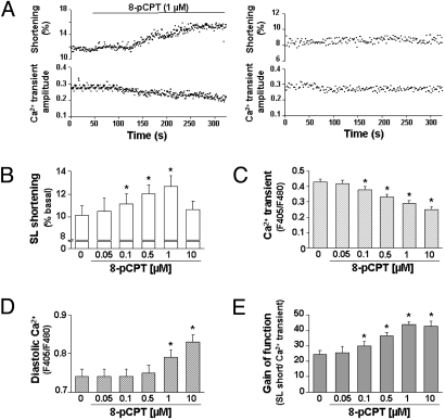
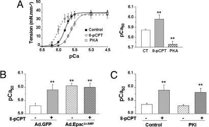
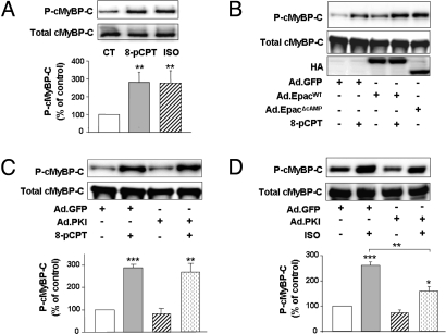
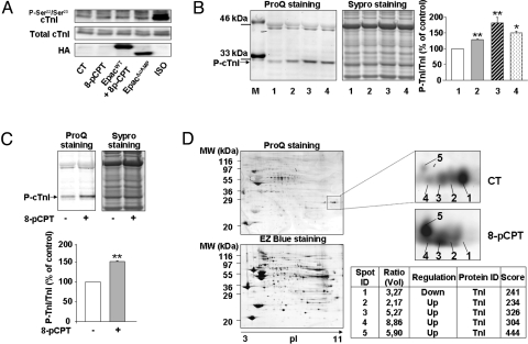
In the heart, cAMP is a key regulator of excitation-contraction coupling and its biological effects are mainly associated with the activity of protein kinase A (PKA). The aim of this study was to investigate the contribution of the cAMP-binding protein Epac (Exchange protein directly activated by cAMP) in the regulation of the contractile properties of rat ventricular cardiac myocytes. We report that both PKA and Epac increased cardiac sarcomere contraction but through opposite mechanisms. Differently from PKA, selective Epac activation by the cAMP analog 8-(4-chlorophenylthio)-2'-O-methyl-cAMP (8-pCPT) reduced Ca(2+) transient amplitude and increased cell shortening in intact cardiomyocytes and myofilament Ca(2+) sensitivity in permeabilized cardiomyocytes. Moreover, ventricular myocytes, which were infected in vivo with a constitutively active form of Epac, showed enhanced myofilament Ca(2+) sensitivity compared to control cells infected with green fluorescent protein (GFP) alone. At the molecular level, Epac increased phosphorylation of 2 key sarcomeric proteins, cardiac Troponin I (cTnI) and cardiac Myosin Binding Protein-C (cMyBP-C). The effects of Epac activation on myofilament Ca(2+) sensitivity and on cTnI and cMyBP-C phosphorylation were independent of PKA and were blocked by protein kinase C (PKC) and Ca(2+) calmodulin kinase II (CaMKII) inhibitors. Altogether these findings identify Epac as a new regulator of myofilament function.


Conflict of interest statement

The authors declare no conflict of interest.

Figures

Fig. 1.
Fig. 1.
Epac regulates the contractile machinery. (A) Effect of 8-pCPT (1 μM) perfusion on sarcomere shortening (Left, Upper) and intracellular calcium transient amplitude (Left, Lower) in intact ventricular cardiomyocytes stimulated at 1 Hz. Activation of Epac increased progressively SL shortening and reduced the amplitude of Ca2+ transient. The maximal effect was observed within 5 min. Right, a time control of SL shortening and calcium transient. (B–E) Concentration-dependent effect of 8-pCPT on SL shortening (B), Ca2+ transient amplitude (C), diastolic calcium level (D), and the gain of function (E), which corresponds to the ratio between SL shortening and calcium transient amplitudes (n = 11 cells). *, P < 0.05 versus control.
Fig. 2.
Fig. 2.
Epac regulates myofilament Ca2+ sensitivity in a PKA-independent manner. (A) The relationship between Ca2+-activated tension and intracellular Ca2+ content was measured in isolated, permeabilized cardiomyocytes at 2.3 μm SL. The relationship was fitted with a modified Hill equation and the pCa at which half of the maximal tension is developed (pCa50) was determined as an index of myofilament Ca2+ sensitivity (see SI Materials and Methods for more details). Preincubation of cells with 8-pCPT (1 μM for 10 min) increased myofilament Ca2+ sensitivity as indicated by the shift toward the left of the curve and the increase in pCa50 (n = 14 cells). In similar conditions, recombinant PKA catalytic subunit induced an opposite effect that reflects desensitization of the myofilaments (n = 10 cells). (B) Constitutively active Epac (EpacΔcAMP) increased myofilament Ca2+ sensitivity as indexed by pCa50. The left ventricular free wall of rats was infected with adenoviruses encoding GFP (Ad-GFP) (control) or bicistronic adenoviruses coexpressing GFP and EpacΔcAMP (Ad-EpacΔcAMP). Three days later, cells were isolated and myofilament Ca2+ sensitivity of permeabilized ventricular cardiomyocytes expressing GFP was measured after incubation, or not, with 8-pCPT (1 μM) for 10 min. Ad-GFP, n = 10; Ad-EpacΔcAMP, n = 16 cells (3 rats per condition). (C) Effect of PKI, a PKA inhibitor, on 8-pCPT-induced myofilament Ca2+ sensitization. Myofilament Ca2+ sensitivity (pCa50) was determined in permeabilized adult ventricular cardiomyocytes treated, or not, with 8-pCPT (1 μM) for 10 min and in the presence or absence of PKI (5 μM); n = 16 cells (3 rats). Results are expressed as means ± SEM. **, P < 0.01 compared with nontreated cells.
Fig. 3.
Fig. 3.
Epac regulates phosphorylation of the thick filament protein cMyBP-C. (A) Effect of 8-pCPT (1 μM) or ISO (100 nM) on cMyBP-C phosphorylation (P-cMyBP-C) in freshly isolated adult cardiomyocytes. Cells were incubated with the drugs for 10 min and P-cMyBP-C was determined by Western blotting using an anti-P-cMyBP-C (P-Ser282) antibody as described in SI Materials and Methods. Membranes were then stripped and probed for total cMyBP-C expression to confirm equivalent protein loading. Lower, immunoblots were quantified and data were normalized to total cMyBP-C expression. (B and C) Effect of 8-pCPT (1 μM) and (D) effect of ISO (100 nM) on P-cMyBP-C in adult cardiomyocytes infected with Ad-GFP, Ad-EpacWT, Ad-EpacΔcAMP, or Ad-PKI (PKA inhibitor). Immunoblots against P-cMyBP-C and total cMyBP-C were performed as in A. (B) An anti-HA antibody was used to monitor the expression levels of EpacWT and EpacΔcAMP. (C and D) Immunoblots were quantified and data normalized to total cMyBP-C. Results are means ± SEM from 6 (A) or 4 (B–D) independent experiments. *, P < 0.05; **, P < 0.01; ***, P < 0.001 compared with control or indicated values. CT, control.
Fig. 4.
Fig. 4.
Epac regulates phosphorylation of cTnI in a PKA-independent manner. (A) Representative Western blot showing cTnI phosphorylation (P-cTnI) at PKA sites (P-Ser22/P-Ser23) (Top). Isolated adult cardiac myocytes were infected with control Ad-GFP (CT), Ad-EpacWT, or Ad-EpacΔcAMP for 36 h. Cells were then treated, or not, with 1 μM 8-pCPT or 100 nM ISO for 10 min and P-Ser22/P-Ser23 cTnI was determined by Western blotting as described in SI Materials and Methods. Total cTnI expression is shown (Middle), and anti-HA antibody was used to monitor the expression levels of EpacWT and EpacΔcAMP (Bottom). (B) Adult cardiac myocytes were infected for 36 h with control Ad-GFP (CT, lane 1), Ad-GFP treated with 1 μM 8-pCPT (lane 2), Ad-EpacWT treated with 1 μM 8-pCPT (lane 3), or Ad-EpacΔcAMP (lane 4). (C) Myocytes were infected with Ad-PKI and were then treated or not with 1 μM 8-pCPT for 10 min as in A and B. Protein phosphorylation was visualized with ProQ Diamond phospho-protein gel stain, followed by total protein staining with Sypro Ruby to confirm equal loading. Right, protein phosphorylation was normalized to the total protein content revealed by SYPRO Ruby staining. Results are means ± SEM of 8 (B) or 4 (C) independent experiments. *, P < 0.05; **, P < 0.01 compared with control values. Tandem mass spectrometry revealed that the phosphorylated 23-kDa band corresponded to cTnI. (D) ProQ Diamond and EZ Coomassie Blue staining of a representative 2D gel. Isolated cardiomyocytes were incubated in the absence or the presence of 8-pCPT (1 μM, 10 min). Protein extracts were separated by 2D electrophoresis using isoelectric focusing strips (18 cm, pH 3–11, nonlinear). A representative enlargement of the gel showing the marked region is illustrated. Five spots were excised and analyzed by LC/MS/MS to show the presence of cTnI. Densitometric analysis of intensities of protein spots was performed and ratios of volume values are indicated in the table. Changes in intensities of protein spots are indicated as increased (up) or decreased (down) in the stimulated 8-pCPT vs. control (CT). The 5 protein spots were identified using the MASCOT Search Engine. The score refers to the degree of similarity between a sample and a searched database match. A score of ≥65 is considered a properly identified match. ID, identity.
Fig. 5.
Fig. 5.
Epac signaling pathways involved in myofilament protein regulation. (A) Expression analysis of Epac and its potential downstream effectors in nonpermeabilized (intact) and permeabilized cardiac myocytes. The indicated proteins were revealed by Western blotting as described in SI Materials and Methods. (B) Shift of myofilament Ca2+ sensitivity of activation (ΔpCa50) induced by 8-pCPT in permeabilized cardiomyocytes preincubated with a PLC inhibitor (U73122, 5 μM), a PKC inhibitor (Calphostin C, 0.5 μM) or a CaMKII inhibitor (KN-93, 2 μM). Average values were expressed as the difference in pCa50 between nonstimulated (no inhibitor) and 8-pCPT-treated cells (n = 8–13 cells per condition). (C and E) Effect of a PKC inhibitor, calphostin-C (0.5 μM), and (D and F) effect of a CaMKII inhibitor, KN-93 (0.5 μM), on P-cTnI and P-cMyBP-C in isolated cardiomyocytes. In C and D phosphorylation of the 23-kDa band corresponding to P-cTnI was revealed by ProQ Diamond phosphoprotein gel staining, followed by total protein staining with Sypro Ruby to confirm equal loading. Representative gels are shown of 4 independent experiments. (E and F) Western blots were performed to probe for P-cMyBP-C and total cMyBP-C expression. Lower, immunoblots were quantified and data normalized to total cMyBP-C expression. Results are means ± SEM from 5 (E) and 4 (F) independent experiments. *, P < 0.05; ***, P < 0.001 compared with control and indicated values.

Similar articles

Cited by

References

    1. Bers DM. Cardiac excitation-contraction coupling. Nature. 2002;415(6868):198–205. - PubMed
    1. Solaro RJ. Modulation of cardiac myofilament activity by protein phosphorylation. In: Page E, Fozzard H, Solaro RJ, editors. Handbook of Physiology. New York: Oxford Univ Press; 2001. pp. 264–300.
    1. Layland J, Solaro RJ, Shah AM. Regulation of cardiac contractile function by troponin I phosphorylation. Cardiovasc Res. 2005;66(1):12–21. - PubMed
    1. Kunst G, et al. Myosin binding protein C, a phosphorylation-dependent force regulator in muscle that controls the attachment of myosin heads by its interaction with myosin S2. Circ Res. 2000;86(1):51–58. - PubMed
    1. Cazorla O, et al. Length and protein kinase A modulations of myocytes in cardiac myosin binding protein C-deficient mice. Cardiovasc Res. 2006;69(2):370–380. - PubMed

Publication types

MeSH terms