. 2009 Aug 18;106(33):13731-6.
doi: 10.1073/pnas.0906509106. Epub 2009 Jul 31.

The repeat domain of the melanosome fibril protein Pmel17 forms the amyloid core promoting melanin synthesis

Affiliations

The repeat domain of the melanosome fibril protein Pmel17 forms the amyloid core promoting melanin synthesis

Ryan P McGlinchey et al. Proc Natl Acad Sci U S A. .

Abstract

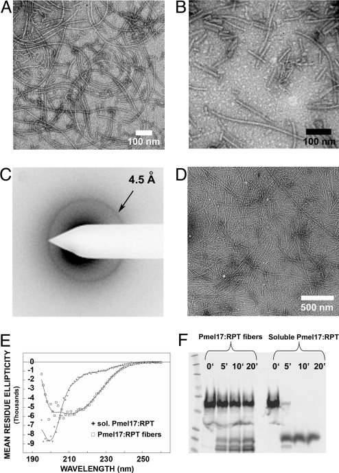
Pmel17 is a melanocyte protein necessary for eumelanin deposition 1 in mammals and found in melanosomes in a filamentous form. The luminal part of human Pmel17 includes a region (RPT) with 10 copies of a partial repeat sequence, pt.e.gttp.qv., known to be essential in vivo for filament formation. We show that this RPT region readily forms amyloid in vitro, but only under the mildly acidic conditions typical of the lysosome-like melanosome lumen, and the filaments quickly become soluble at neutral pH. Under the same mildly acidic conditions, the Pmel filaments promote eumelanin formation. Electron diffraction, circular dichroism, and solid-state NMR studies of Pmel17 filaments show that the structure is rich in beta sheet. We suggest that RPT is the amyloid core domain of the Pmel17 filaments so critical for melanin formation.


Conflict of interest statement

The authors declare no conflict of interest.

Figures

Fig. 1.
Fig. 1.
The Pmel RPT domain forms fibers under nondenaturing conditions. (A) Full-length Pmel17 is composed of 10 domains. SIG, signal peptide; NTD, N-terminal domain; PKD, polycystic kidney disease-like domain; RPT, proline/serine/threonine-rich repeat domain; KRG, kringle-like domain; TM, transmembrane domain; CTD, C-terminal domain; GAP1, GAP2, and GAP3, undefined domains. Known N-glycosylation sites are indicated above the NTD, RPT, and GAP3 domains by a “Y”. The RPT domain is O-glycosylated. The fragments examined for amyloid formation are shown with endpoints and His6 tag, if any. (B) SDS/PAGE (10%) analysis of RPT purified under denaturing conditions. (C) Sequence of the RPT domain, showing the imperfect repeats rich in proline, serine and threonine residues. (D) Transmission electron micrographs of Pmel17:RPT fibers negatively stained with uranyl acetate.
Fig. 2.
Fig. 2.
pH stability of RPT fibers. (A) Transmission electron micrograph images showing pH stability between pH 4.0–9.0 of RPT fibers formed at pH 5.0. (B) Comparison of ThT fluorescence of RPT with that of Ure2p, Sup35NM, and HET-s at pH 5.0. (C) RPT fibers monitored by thioflavin T over time at pH 5.0–8.0. (D) RPT fibers monitored by thioflavin T over time at pH 5.0–8.0.
Fig. 3.
Fig. 3.
RPT fibrils prepared by seeding or agitation. (A) Fibrils made by seeding with 5% RPT seeds for 2 weeks. (B) Fibrils made by 1 week incubation with gentle agitation. (C) Electron diffraction of RPT fibers formed in vitro exhibits a reflection at 4.5 ± 0.2 Å indicative of a β sheet structure. (D) Electron micrograph of sample used in C. (E) Circular dichroism spectra of soluble and fibrous RPT support a β-sheet rich structure of the fibrils. Based on curve-fitting, RPT is 6 ± 1% α-helix, 47 ± 2% β-sheet, 19 ± 1% β-turn, and 28 ± 1% remainder. (F) Equal concentrations of soluble and fibrous RPT exposed to Proteinase K support a protease-resistant core in the fiber form.
Fig. 4.
Fig. 4.
Amyloid of recombinant RPT accelerates melanin synthesis. (Upper) A time course of melanin synthesis in vitro shows that RPT fibers enhance melanin formation per unit time when compared to the soluble form. (Lower) Comparison of melanin synthesis facilitated by amyloid of RPT (q) with that of Sup35NM (p) and HET-s (l). Reactions were performed in duplicate.

References

    1. Barnhart MM, Chapman MR. Curli biogenesis and function. Annu Rev Microbiol. 2006;60:131–147. - PMC - PubMed
    1. Sunde M, Kwan AH, Templeton MD, Beever RE, MacKay JP. Structural analysis of hydrophobins. Micron. 2008;39:773–784. - PubMed
    1. Podrabsky JE, Carpenter JF, Hand SC. Survival of water stress in annual fish embryos: Dehydration avoidance and egg amyloid fibers. Am J Physiol Regulatory Integrative Comp Physiol. 2001;280:R123–R131. - PubMed
    1. Iconomidou VA, Vriend G, Hamodrakas SJ. Amyloids protect the sildmoth oocyte and embryo. FEBS Lett. 2000;479:141–145. - PubMed
    1. Benkemoun L, Saupe SJ. Prion proteins as genetic material in fungi. Fungal Genet Biol. 2006;43:789–803. - PubMed

Publication types

LinkOut - more resources