. 2009 Aug 18;106(33):13713-8.
doi: 10.1073/pnas.0812374106. Epub 2009 Aug 4.

Ca2+ binding by domain 2 plays a critical role in the activation and stabilization of gelsolin

Affiliations

Ca2+ binding by domain 2 plays a critical role in the activation and stabilization of gelsolin

Shalini Nag et al. Proc Natl Acad Sci U S A. .

Abstract

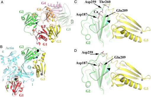
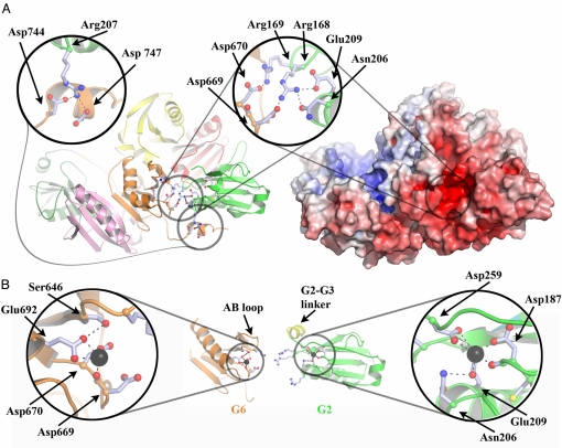
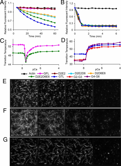
Gelsolin consists of six homologous domains (G1-G6), each containing a conserved Ca-binding site. Occupation of a subset of these sites enables gelsolin to sever and cap actin filaments in a Ca-dependent manner. Here, we present the structures of Ca-free human gelsolin and of Ca-bound human G1-G3 in a complex with actin. These structures closely resemble those determined previously for equine gelsolin. However, the G2 Ca-binding site is occupied in the human G1-G3/actin structure, whereas it is vacant in the equine version. In-depth comparison of the Ca-free and Ca-activated, actin-bound human gelsolin structures suggests G2 and G6 to be cooperative in binding Ca(2+) and responsible for opening the G2-G6 latch to expose the F-actin-binding site on G2. Mutational analysis of the G2 and G6 Ca-binding sites demonstrates their interdependence in maintaining the compact structure in the absence of calcium. Examination of Ca binding by G2 in human G1-G3/actin reveals that the Ca(2+) locks the G2-G3 interface. Thermal denaturation studies of G2-G3 indicate that Ca binding stabilizes this fragment, driving it into the active conformation. The G2 Ca-binding site is mutated in gelsolin from familial amyloidosis (Finnish-type) patients. This disease initially proceeds through protease cleavage of G2, ultimately to produce a fragment that forms amyloid fibrils. The data presented here support a mechanism whereby the loss of Ca binding by G2 prolongs the lifetime of partially activated, intermediate conformations in which the protease cleavage site is exposed.


Conflict of interest statement

The authors declare no conflict of interest.

Figures

Fig. 1.
Fig. 1.
Structures of Ca-free human gelsolin and human G1–G3/actin. (A) Schematic representation of the structure of Ca-free human gelsolin. The arrowhead in this part, as in the others, points toward the peptide bond between Arg-172 and Ala-173, which gets cleaved in FAF. (B) The structure of human G1–G3 bound to actin. G2 is shown in a similar orientation as in A. There are five Ca2+ ions (black spheres) associated with this structure, one bound to each gelsolin domain, another sandwiched between G1 and actin, and one at the ATP-binding site of actin. (C) Close up of the Ca-coordinating residues in G2 from human G1–G3/actin. Only G2–G3 is shown for clarity. (D) Close up of the vacant G2 Ca-binding site from equine G1–G3/actin (Protein Data Bank ID 1RGI). Protein representations were generated here and in the figures that follow by using PYMOL (http://pymol.sourceforge.net/).
Fig. 2.
Fig. 2.
Structural interdependence of the G2 and G6 Ca-binding sites. (A) Schematic and electrostatic surface representations of Ca-free human gelsolin, highlighting the charged residues at the G2–G6 interface. (B) Schematic representations of Ca-bound G6 and G2, taken from the structures of Ca-bound equine G4–G6/actin (Protein Data Bank ID 1H1V) and human G1–G3/actin, respectively, in orientations similar to those presented in A. G2 and G6 have been translated relative to their positions in A to avoid steric clashes. Note that the Ca ions are coordinated by residues that previously made up the network of interactions between G2 and G6 in A.
Fig. 3.
Fig. 3.
Ca effects on gelsolin structure and function. (A and B) Actin depolymerization assays. A total of 6 μM of each protein was added to 12 μM F actin in the presence of (A) 1 mM EGTA and (B) 10 μM free Ca2+. Single mutants (D2, E2, D6, and E6) and double mutants (D2D6, D2E6, E2D6, E2E6, and D6E6) were indistinguishable from GFL and are not shown for clarity. TIRF analysis confirmed that gelsolin severs actin under these conditions (Movie S4). (C and D) Thermal shift assays. (C) Effect of Ca concentration on thermal stability of GFL and the D2E2D6E6 quadruple mutant. (D) Effect of Ca concentration on thermal stability of GTL, G2–G3, and G4–G6. pCa refers to the theoretical free Ca concentration that is not bound to EGTA in the absence of protein. (E–G) TIRF assay. TIRF images of actin filaments (500 nM) severed by (E) GTL (2.5 nM), (F) D2E2D6E6 (2.5 nM), and (G) GFL (2.5 nM) in 1 mM EGTA buffer. Shown are 40 × 40 μm snapshots at four different time points (from left to right): (E) before mixing and 40 s, 80 s, and 17.5 min after mixing; (F) before mixing and 2, 4, and 17.5 min after mixing; and (G) before mixing, 140 s after mixing, 4 mins after mixing, and 35 s after adding Ca2+ (5 mM) to the stable mixture. Movies of E, F, and G and a description of the TIRF experiment are available in the SI Text and Movies S1–S3.

Similar articles

Cited by

References

    1. Silacci P, et al. Gelsolin superfamily proteins: Key regulators of cellular functions. Cell Mol Life Sci. 2004;61:2614–2623. - PMC - PubMed
    1. Yin HL, Kwiatkowski DJ, Mole JE, Cole FS. Structure and biosynthesis of cytoplasmic and secreted variants of gelsolin. J Biol Chem. 1984;259:5271–5276. - PubMed
    1. Kwiatkowski DJ, Mehl R, Yin HL. Genomic organization and biosynthesis of secreted and cytoplasmic forms of gelsolin. J Cell Biol. 1988;106:375–384. - PMC - PubMed
    1. Way M, Weeds A. Nucleotide sequence of pig plasma gelsolin. J Mol Biol. 1988;203:1127–1133. - PubMed
    1. Koepf EK, Hewitt J, Vo H, MacGillivray RTA, Burtnick LD. Equus caballus gelsolin cDNA sequence and protein structural implications. Eur J Biochem. 1998;251:613–621. - PubMed

Publication types