. 2009 Aug 18;106(33):13939-44.
doi: 10.1073/pnas.0907143106. Epub 2009 Aug 4.

Cell-type-specific isolation of ribosome-associated mRNA from complex tissues

Affiliations

Cell-type-specific isolation of ribosome-associated mRNA from complex tissues

Elisenda Sanz et al. Proc Natl Acad Sci U S A. .

Abstract

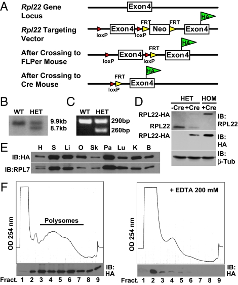
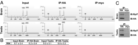
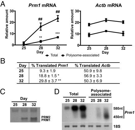
Gene profiling techniques allow the assay of transcripts from organs, tissues, and cells with an unprecedented level of coverage. However, most of these approaches are still limited by the fact that organs and tissues are composed of multiple cell types that are each unique in their patterns of gene expression. To identify the transcriptome from a single cell type in a complex tissue, investigators have relied upon physical methods to separate cell types or in situ hybridization and immunohistochemistry. Here, we describe a strategy to rapidly and efficiently isolate ribosome-associated mRNA transcripts from any cell type in vivo. We have created a mouse line, called RiboTag, which carries an Rpl22 allele with a floxed wild-type C-terminal exon followed by an identical C-terminal exon that has three copies of the hemagglutinin (HA) epitope inserted before the stop codon. When the RiboTag mouse is crossed to a cell-type-specific Cre recombinase-expressing mouse, Cre recombinase activates the expression of epitope-tagged ribosomal protein RPL22(HA), which is incorporated into actively translating polyribosomes. Immunoprecipitation of polysomes with a monoclonal antibody against HA yields ribosome-associated mRNA transcripts from specific cell types. We demonstrate the application of this technique in brain using neuron-specific Cre recombinase-expressing mice and in testis using a Sertoli cell Cre recombinase-expressing mouse.


Conflict of interest statement

The authors declare no conflict of interest.

Figures

Fig. 1.
Fig. 1.
Targeting the Rpl22 genomic locus. (A) Targeting strategy for the genomic locus of Rpl22. A loxP site was inserted 5′ to the wild-type exon 4. A loxP-FRT-neomycin resistance-FRT cassette was inserted 3′ to the wild-type exon 4 followed by a modified Rpl22 exon 4 containing the HA epitope tag inserted before the Rpl22 stop codon. F1 heterozygous offspring were bred to FLPeR mice to remove the neomycin cassette used for selection in the ES cells. Crossing the RiboTag mouse to a Cre recombinase-expressing mouse results in deletion of the wild-type exon 4 in the target cell population and replacement with the Rpl22HA exon 4. (B) Southern blot strategy used to identify correctly targeted ES cells. The wild-type HindIII/SfiI DNA fragment is 9,908 bp, while the correctly targeted HindIII/SfiI fragment is 8,749 bp. (C) PCR products using oligonucleotides that amplify the loxP-containing intron sequence 5′ to the wild-type exon 4 of the Rpl22 gene. The wild-type PCR product is 260 bp, while the mutant PCR product is 290 bp. (D) Western analysis of wild-type RPL22 or RPL22ha in a control Meox2+/+:Rpl22ha/+ mouse, a double heterozygote Meox2Cre/+:Rpl22ha/+ mouse, and a Rpl22ha-expressing homozygous mouse. Blots were probed with anti-RPL22 and anti-HA antibodies. RPL22ha results in a 23-kDa protein, while the native RPL22 is a 15-kDa protein. (E) Western blot using tissue homogenates from a Rpl22ha-expressing homozygous mouse. The blot was probed with anti-HA antibody and then reprobed with anti-RPL7 antibody. H, heart; S, spleen; Li, liver; O, ovary; Sk, skeletal muscle; Pa, pancreas; Lu, lung; K, kidney; B, brain. (F) A254 absorbance profile of 15% to 50% sucrose density gradients from a Rpl22ha-expressing homozygous mouse brain in the absence (left) or presence (right) of 200 mM EDTA. Western blot analysis (below each profile) of trichloroacetic acid-precipitated fractions probed with anti-HA antibody.
Fig. 2.
Fig. 2.
A cartoon that outlines the RiboTag methodology. The RiboTag mouse is crossed to any available Cre recombinase-expressing mouse line, which results in the deletion of wild-type exon 4 and replacement with the HA-tagged exon 4 only in cells that express Cre recombinase. The cells within the tissue or organ that now express RPL22ha-tagged ribosomes are homogenized, and HA antibody-coupled magnetic beads are added to the cleared homogenate. After an overnight incubation at 4 °C, the magnetic beads that have immunoadsorbed polysomes are washed with a high salt buffer before extraction of the mRNA transcripts, which can then be analyzed by qRT-PCR, microarray, or RNA-seq.
Fig. 3.
Fig. 3.
Immunoprecipitation of polysomes from Rpl22ha-expressing homozygous mouse brain and testis. (A) Agilent Technologies 2100 Bioanalyzer electropherogram analysis of total RNA from brain and testis immunoprecipitates. (B) RIN values from Agilent Technologies 2100 Bioanalyzer analysis demonstrating recovery of high quality RNA from the pellets (RIN values >8.0). (C) Western blots using anti-HA antibody and anti-RPL7 antibody demonstrating the presence of RPL7 (a component of the large subunit of the ribosome) specifically in anti-HA versus anti-Myc pellets.
Fig. 4.
Fig. 4.
Enrichment of transcripts expressed in dopaminergic neurons (DAT-Cre:RiboTag), medium spiny neurons of the striatum (DARPP32-Cre:RiboTag), and Sertoli cells of the testis (AMH-Cre:RiboTag) using the RiboTag technology. (A) Immunohistochemistry on 25-μm brain and testis sections using anti-HA antibody and marker antibodies to proteins known to be expressed in the cell type or area under study. (Top panels) Immunohistochemistry on DAT-Cre:RiboTag mouse brain. Anti-HA staining is shown in green and anti-tyrosine hydroxylase (TH) staining is shown in red. Merged image shows the presence of both TH and HA staining in the same cell types. (Middle panels) Immunohistochemistry on DARPP32-Cre:RiboTag mouse brain. Anti-HA staining is shown in green and anti-TH staining is shown in red. TH was used as a striatal marker. Merged image shows striatal localization of the HA staining. (Bottom panels) Immunohistochemistry on AMH-Cre:RiboTag mouse testis. Anti-HA staining is shown in green and TO-PRO-3 (Invitrogen) nuclear staining is shown in blue. (B) Anti-HA Western blots using 10% weight per volume homogenates of DAT-Cre:RiboTag brain, DARPP32-Cre:RiboTag brain, and AMH-Cre:RiboTag testis demonstrating varying levels of RPL22ha protein depending on the Cre recombinase-expressing mouse line used. (C) Western blots after immunoprecipitation of brain and testis homogenates using anti-HA or control anti-Myc antibodies and demonstrating specific immunoprecipitation of RPL22ha protein in HA versus Myc pellets. Two percent of input (I) and supernatant (S) samples, and 4% of pellet (P) samples were loaded on the gel. (D) qRT-PCR analysis of transcripts expressed in dopaminergic neurons, medium spiny neurons of the striatum, and Sertoli cells of the testis after immunoprecipitation of polysomes from DAT-Cre:RiboTag brain, DARPP32-Cre:RiboTag brain, and AMH-Cre:RiboTag testis. Total RNA was isolated from input and from anti-HA or control anti-Myc pellets after overnight immunoprecipitation of polysomes from 10% weight per volume homogenates of brain or testis. All cell-specific marker genes and control genes not expressed in the target cells were normalized to beta-actin (Actb) levels using the ΔΔCt method. The immunoprecipitated RNA samples were compared to the input sample in each case. The transcripts analyzed by qRT-PCR were: Tyrosine hydroxylase (Th); dopamine transporter (DAT, Slc6a3); 2′,3′-cyclic nucleotide 3′ phosphodiesterase (CNPase, Cnp); dopamine- and cAMP-regulated phosphoprotein (DARPP-32, Ppp1r1b); prodynorphin (Pdyn); proenkephalin (Penk1); follicle-stimulating hormone receptor (Fshr); transferrin (Trf); inhibin beta-B (Inhbb); protamine 2 (Prm2). ***, P < 0.001; **, P < 0.01. Values are the mean ± SEM of three independent experiments.
Fig. 5.
Fig. 5.
Immunoprecipitation of actively translated Prm1 mRNA from Rpl22ha-expressing homozygous mouse testis. (A) qRT-PCR Taqman assay using a probe specific for Prm1 mRNA comparing input (total RNA) and HA-immunoprecipitated (polysome-associated RNA) from P25, P28, and P32 Rpl22ha-expressing homozygous mouse testis homogenates. qRT-PCR analysis of beta-actin (Actb) transcripts was performed as a control. Values are the mean ± SEM of three independent experiments. (B) Table showing the percentage of polysome-associated transcripts compared to total transcripts (% Translated) of Prm1 and Actb at each time point. (C) Coomassie Brilliant Blue staining showing basic proteins isolated from nuclear pellets of P25, P28, and P32 Rpl22ha-expressing homozygous mouse testis. Samples were resolved on 15% acrylamide acid-urea gels. (D) Northern blot analysis revealing the predominance of partially deadenylated Prm1 transcripts in the polysome-associated samples.

Similar articles

Cited by

References

    1. Arlotta P, et al. Neuronal subtype-specific genes that control corticospinal motor neuron development in vivo. Neuron. 2005;45:207–221. - PubMed
    1. Buchstaller J, et al. Efficient isolation and gene expression profiling of small numbers of neural crest stem cells and developing Schwann cells. J Neurosci. 2004;24:2357–2365. - PMC - PubMed
    1. Hempel CM, Sugino K, Nelson SB. A manual method for the purification of fluorescently labeled neurons from the mammalian brain. Nat Protoc. 2007;2:2924–2929. - PubMed
    1. Lobo MK, et al. FACS-array profiling of striatal projection neuron subtypes in juvenile and adult mouse brains. Nat Neurosci. 2006;9:443–452. - PubMed
    1. Rossner MJ, et al. Global transcriptome analysis of genetically identified neurons in the adult cortex. J Neurosci. 2006;26:9956–9966. - PMC - PubMed

Publication types

LinkOut - more resources