. 2009 Aug 18;106(33):13933-8.
doi: 10.1073/pnas.0904744106. Epub 2009 Aug 4.

GATA6 mutations cause human cardiac outflow tract defects by disrupting semaphorin-plexin signaling

Affiliations

GATA6 mutations cause human cardiac outflow tract defects by disrupting semaphorin-plexin signaling

Kazuki Kodo et al. Proc Natl Acad Sci U S A. .

Abstract

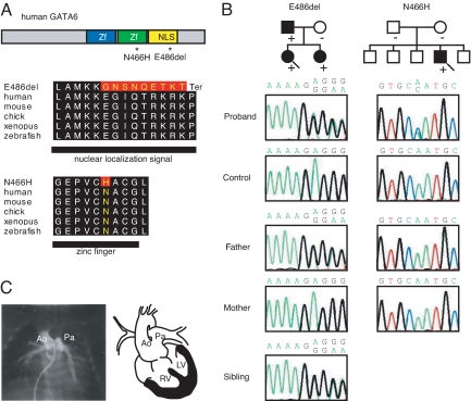
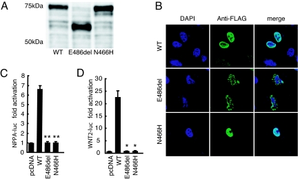
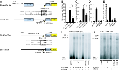
Congenital heart diseases (CHD) occur in nearly 1% of all live births and are the major cause of infant mortality and morbidity. Although an improved understanding of the genetic causes of CHD would provide insight into the underlying pathobiology, the genetic etiology of most CHD remains unknown. Here we show that mutations in the gene encoding the transcription factor GATA6 cause CHD characteristic of a severe form of cardiac outflow tract (OFT) defect, namely persistent truncus arteriosus (PTA). Two different GATA6 mutations were identified by systematic genetic analysis using DNA from patients with PTA. Genes encoding the neurovascular guiding molecule semaphorin 3C (SEMA3C) and its receptor plexin A2 (PLXNA2) appear to be regulated directly by GATA6, and both GATA6 mutant proteins failed to transactivate these genes. Transgenic analysis further suggests that, in the developing heart, the expression of SEMA3C in the OFT/subpulmonary myocardium and PLXNA2 in the cardiac neural crest contributing to the OFT is dependent on GATA transcription factors. Together, our data implicate mutations in GATA6 as genetic causes of CHD involving OFT development, as a result of the disruption of the direct regulation of semaphorin-plexin signaling.


Conflict of interest statement

The authors declare no conflict of interest.

Figures

Fig. 1.
Fig. 1.
Identification of GATA6 mutations in patients with persistent truncus arteriosus (PTA). (A) The structure of the human GATA6 gene, the position of the two mutations (asterisks), and conservation of alignment between species are shown. Zf, zinc finger; NLS, nuclear localization signal. Changes in amino acids are highlighted in red. The E486del mutation causes two nucleotide deletions, resulting in nine amino acid changes followed by P489 to termination (Ter) codon. (B) Pedigree indicating cardiac phenotype and the presence (+) or absence (-) of the GATA6 mutation in the family of proband A (E486del; arrow) and proband B (N466H; arrow). The probands are indicated by arrows. (circle), female; (box), male; (solid fill), with CHD. In addition, a sequence chromatogram of one frame shift mutation (E486del, proband A) and a point mutation (N466H, proband B) are shown. (C) A catheter angiogram of proband A reveals a single common outflow tract with no septation between the aorta (Ao) and pulmonary artery (Pa), diagnosed as PTA. A schematic diagram of PTA is shown on the right. RV, right ventricle; LV, left ventricle.
Fig. 2.
Fig. 2.
Molecular size, nuclear localization and transactivation ability of GATA6 mutant proteins. (A) Western blot analyses show that the band of the FLAG-tagged E486del mutant GATA6 protein is approximately 15 kDa smaller than the FLAG-tagged wild-type (WT) and N466H mutant proteins. (B) Nuclear localization of FLAG-tagged GATA6 wild-type (WT) and mutant (E486del and N466H) proteins, shown in green. Purple staining (DAPI) indicates the nucleus. The E486del mutant shows an abnormal localization pattern. (C and D) Relative luciferase activity in HeLa cells transfected with wild-type GATA6 (WT) or GATA6 mutant (E486del or N466H) expression constructs and NPPA-luc (C) or WNT2-luc (D). Both mutants are unable to activate the transcription of each reporter construct (C: WT vs. E486del, P = 0.0023; WT vs. N466H, P = 0.0016, n = 3; D: E486del, P = 0.013; N466H, P = 0.013; two-tailed unpaired t test, n = 3). *, P < 0.05; **, P < 0.01 compared with WT.
Fig. 3.
Fig. 3.
GATA6 mutations attenuate the direct regulation of semaphorin 3C (SEMA3C) and its receptor plexin A2 (PLXNA2) by GATA6. (A) Structure of SEMA3C-luc and PLXNA2-luc with a consensus GATA-binding site (Site1 and Site2). and structure of SEMA3C-luc with site-directed mutagenesis for Site1 (ΔSite1-luc) and PLXNA2-luc with site-directed mutagenesis for Site2 (ΔSite2-luc) are shown. (B and C) Fold increase in relative luciferase activity directed by SEMA3C promoter-luc constructs or PLXNA2 promoter-luc constructs in HeLa cells cotransfected with GATA6-pcDNA. The Site1 or the Site2 mutation results in a significant decrease in activation by GATA6 compared with controls (B; SEMA3C-luc vs. ΔSite1-luc, P = 0.027; n = 3; C; PLXNA2-luc vs. ΔSite2-luc, P = 0.037, n = 5). *, P < 0.05. (D and E) Relative luciferase activity in HeLa cells transfected with pcDNA, GATA6 wild-type (WT) or each mutant (E486del or N466H) expression construct and SEMA3C-luc or PLXNA2-luc. Both mutants display a significant decrease in activation of each promoter compared with WT (D; E486del, P = 0.0028; N466H, P = 0.0030; two-tailed unpaired t test, n = 4; E; E486del, P = 0.0031; N466H, P = 0.0030, n = 3). **, P < 0.01 compared with WT. (F) Binding of each GATA6 protein for Site1. GATA6 WT can bind Site1, but the E486del and N466H mutants cannot. (G) Binding of each GATA6 protein for Site2. GATA6 WT and N466H mutant can bind Site2, but the E486del mutant cannot. SS, super shift.
Fig. 4.
Fig. 4.
GATA cis-elements in the Sema3c and Plxna2 enhancer/promoter are essential for their expression in outflow tract development. (A) Schematic diagram of the mouse 4.7-kb Sema3c promoter-lacZ reporter plasmid with or without site-directed mutagenesis of Site1 (ΔSite1-lacZ). The number of transgenic embryos analyzed is indicated. (B and C) Embryonic day (E) 11.5 embryos harboring the Sema3c-lacZ transgene. (D and E) Clearing of the embryo revealed lacZ-positive cells migrating into the outflow tract (oft) and subpulmonary myocardium (arrowheads), and the right ventricle (rv) shown in the higher-magnification image of the heart. (F and G) E11.5 embryo harboring the ΔSite1-lacZ transgene. (H and I) Clearing of the embryo showed no lacZ-positive cells in the oft and rv, shown in the higher-magnification image of the heart. (J) Schematic diagram of a mouse 1-kb Plxna2 promoter-lacZ reporter plasmid with or without site-directed mutagenesis of Site2 (ΔSite2-lacZ). The number of transgenic embryos analyzed is indicated. (K,L) E12.5 embryo harboring the Plxna2-lacZ transgene. (M) Clearing of embryos revealed lacZ-positive cells in the oft and pharyngeal arch arteries (paa). The white arrow indicates the truncus arteriosus. (N) Higher magnification of the heart. The white arrow indicates the truncus arteriosus. (O and P) E12.5 embryo harboring the ΔSite2-lacZ transgene. (Q) Clearing of embryos revealed no lacZ expression in the oft. (R) Higher magnification of the heart. (Scale bars, 1 mm.) sm, somites; lb, limb bud; nt, dorsal neural tube; ra, right atrium; lv, left ventricle; la, left atrium; 3/4, paa 3 and 4; 6, paa 6.

References

    1. Hoffman JI, Kaplan S. The incidence of congenital heart disease. J Am Coll Cardiol. 2002;39:1890–1900. - PubMed
    1. Thom T, et al. Heart disease and stroke statistics–2006 update: A report from the American Heart Association Statistics Committee and Stroke Statistics Subcommittee. Circulation. 2006;14:e85–151. - PubMed
    1. Williams JM, et al. Factors associated with outcomes of persistent truncus arteriosus. J Am Coll Cardiol. 1999;34:545–553. - PubMed
    1. Srivastava D. Making or breaking the heart: From lineage determination to morphogenesis. Cell. 2006;126:1037–1048. - PubMed
    1. Olson EN. Gene regulatory networks in the evolution and development of the heart. Science. 2006;313:1922–1927. - PMC - PubMed

Publication types

MeSH terms