. 2009 Aug 11;106(32):13606-11.
doi: 10.1073/pnas.0906377106. Epub 2009 Jul 28.

TRPA channels distinguish gravity sensing from hearing in Johnston's organ

Affiliations

TRPA channels distinguish gravity sensing from hearing in Johnston's organ

Yishan Sun et al. Proc Natl Acad Sci U S A. .

Abstract

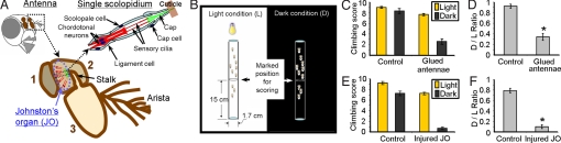
Although many animal species sense gravity for spatial orientation, the molecular bases remain uncertain. Therefore, we studied Drosophila melanogaster, which possess an inherent upward movement against gravity-negative geotaxis. Negative geotaxis requires Johnston's organ, a mechanosensory structure located in the antenna that also detects near-field sound. Because channels of the transient receptor potential (TRP) superfamily can contribute to mechanosensory signaling, we asked whether they are important for negative geotaxis. We identified distinct expression patterns for 5 TRP genes; the TRPV genes nanchung and inactive were present in most Johnston's organ neurons, the TRPN gene nompC and the TRPA gene painless were localized to 2 subpopulations of neurons, and the TRPA gene pyrexia was expressed in cap cells that may interact with the neurons. Likewise, mutating specific TRP genes produced distinct phenotypes, disrupting negative geotaxis (painless and pyrexia), hearing (nompC), or both (nanchung and inactive). Our genetic, physiological and behavioral data indicate that the sensory component of negative geotaxis involves multiple TRP genes. The results also distinguish between different mechanosensory modalities and set the stage for understanding how TRP channels contribute to mechanosensation.


Conflict of interest statement

The authors declare no conflict of interest.

Figures

Fig. 1.
Fig. 1.
Johnston's organ is required for negative geotaxis in Drosophila. (A) Schematic showing the fly head, antenna, Johnston's organ, and a single scolopidium of Johnston's organ. The numbers 1, 2, and 3 refer to the 3 antennal segments. (B) Schematic of the tube-climbing test. In each trial, a group of 10 flies were tapped to the bottom of the tube, and we counted the number of flies crossing the 15-cm threshold line within 15 s as the climbing score. (C) Climbing scores in Light (L) and Dark (D) conditions of control Canton-S (CS) flies (n = 10 trials) and CS flies with the third antennal segment glued to head (n = 12 trials). (D) Ratios of climbing scores (D/L Ratios) for flies in C. (E) Climbing scores of control w1118-WLS flies (n = 10 trials) and w1118-WLS flies with injured Johnston's organ because of second segment pinching (n = 10 trials). (F) D/L Ratios for flies in E. Data are mean ± SEM. *, P < 0.05 by unpaired t test.
Fig. 2.
Fig. 2.
TRP genes have distinct expression patterns in Johnston's organ. (A) Expression of nan-Gal4, iav-Gal4, nompC-Gal4, painGal4, and pyx-Gal4 in Johnston's organ visualized by UAS-GFP, which labels the cytoplasm (Upper, scale bars, 10 μm, except for pyx-Gal4 where scale bar, 30 μm), and UAS-Nuclear DsRed, which labels cell nuclei (Lower, fluorescent images are overlaid on DIC images, scale bars, 30 μm). In the second antennal segment, all labeling appeared to be in Johnston's organ; nan-Gal4, painGal4 and pyx-Gal4 also labeled some cells in the third segment. White dashed arrows indicate direction from cell body to dendrite and cilium. (B) Confocal reconstruction of Johnston's organ expressing both painGal4 and pyx-Gal4 visualized by UAS-Nuclear DsRed (red). Inset shows a different view of the structure with pain and pyx expressing cell nuclei indicated. D, dorsal; A, anterior; M, medial; P, posterior. (C) Expression of both painGal4 and pyx-Gal4 visualized by UAS-myr-mRFP (red), which labels the plasma membrane. Scolopale rods are stained with Alexa633-phalloidin (blue). Dendritic caps are labeled with transgenic GFP-NompA (green). (Scale bar, 20 μm.) The boxed region highlights 2 painGal4-expressing neurons projecting their cilia to pyx-Gal4-expressing cap cells. Three enlarged views of this region are displayed on the right. Arrows indicate cilia and arrowheads indicate a cluster of cap cells.
Fig. 3.
Fig. 3.
Specific TRP mutants impair negative geotaxis behavior in the tube-climbing test. (A) Climbing scores of TRP mutants. nompCf00642 was backcrossed to the w1118-WLS control strain. pain1, painGal4, and pyx3 were backcrossed to the Canton-S (CS) control strain. nan36a and iav3621 were in their original genetic backgrounds. (B) D/L Ratios for flies in A. w1118-WLS (n = 14 trials) and nompCf00642 (n = 27 trials) were compared with unpaired t test. CS (n = 25 trials) and mutants (pain1, n = 25 trials; painGal4, n = 10 trials; pyx3, n = 9 trials; nan36a, 10 trials; iav3621, n = 10 trials) were compared by ANOVA (α level = 0.05) and post hoc test of Games-Howell. (C and D) pyxDf4 flies (n = 22 trials) had normal behavior, but pyxDf9 (n = 24 trials) flies were defective compared with the w1118-WLS control (n = 15 trials). D/L Ratios were analyzed by ANOVA (α level = 0.05) and post hoc test of Games-Howell. Data are mean ± SEM. *, significant difference from the control.
Fig. 4.
Fig. 4.
Function of pain and pyx in peripheral sensory tissues is key for negative geotaxis. (A) Expression of the dominant negative UAS-pyxFAP driven by pyx-Gal4 disrupted negative geotaxis in the climbing assay. Note that in the 3 genotypes each transgene was in homozygous state. *, significant difference from pyx-Gal4 based on ANOVA (α level = 0.05) and post hoc test of Games Howell (ANOVA). n = 10 trials in each group. (B) pyx-Gal4 did not drive mCD8::GFP reporter expression in the brain or thoracico-abdominal ganglion. CNS tissues were stained with the nc82 antibody to visualize neuropil (red) and anti-GFP antibody to visualize the reporter. (Scale bars, 100 μm.) (C) Expression of UAS-pyxFAP driven by Appl-Gal4 (a pan-neuronal Gal4 driver) had no effect on negative geotaxis. n = 9 trials in each group. (D) In the leg, nuclear DsRed expression driven by iav-Gal4 was only detected in femoral chordotonal organs. Inset shows a cluster of nuclei of chordotonal neurons. (E) iav-Gal4 did not drive mCD8::GFP expression in central neurons, but anti-GFP staining was present in projections of sensory afferents from Johnston's organ and leg chordotonal neurons (green). (Scale bars, 100 μm.) (F) Expression of UAS-pain under the control of iav-Gal4 restored negative geotaxis. *, significant difference from each of the 2 controls, based on ANOVA (α level = 0.05) and post hoc test of Games Howell. n = 39 trials for pain1; iav-Gal4/+, n = 35 trials for pain1; UAS-pain/+, and n = 39 trials for pain1; iav-Gal4/UAS-pain. Data are mean ± SEM.
Fig. 5.
Fig. 5.
Specific TRP mutants show defective antennal nerve responses to rotation. (A) Schematic of the recording apparatus. The recording electrode and fly were both mounted on a rotatable platform. Rotation of the platform changes the orientation of the fly body, but the electrode position remains constant relative to the fly. The recording electrode was positioned between the first and second antennal segment where axons of Johnston's organ fasciculate (Inset). (B–D) Bidirectional 90° pitch (B), roll (C), and yaw (D) all induced transient spiking responses in the antennal nerve of Canton-S (CS) wild-type flies. The response was reproducible with repetitive stimulations. Expanded trace in B shows quantification of the response. We counted the number of spikes with amplitudes greater than 2-fold baseline activity. The threshold is represented with the horizontal line in cyan. The counted spikes are marked with blue dots at their negative peaks and represented by a temporally aligned raster above the trace. Variability in the amplitude of individual spikes suggests that multiple units (neurons) were recorded. (E) Spiking responses to bidirectional 90° pitches were abolished by gluing the third antennal segment to the head to prevent its movement, and the response partially recovered after removing the glue. (F) Sample traces for nan36a, iav3621, pain1, painGal4, and pyx3 mutants in response to bidirectional 90° pitches. Each trace represents an example that had a spike number (forward pitch) at the median for all specimens of that genotype. (G) Quantification of spiking responses in the mutants. For nan36a, iav3621, and painGal4. *, significant difference from CS by ANOVA (α level = 0.05) and post hoc test of Games-Howell. *, P < 0.05 by unpaired t test for pyx3. The number (n) of antennae recorded in each group is shown. Data are mean ± SEM.
Fig. 6.
Fig. 6.
The nompC but not the pain and pyx mutations disrupted auditory responses. (A) Schematic of the auditory recording method. Near-field sound, mimicking the Drosophila courtship song was delivered to the antenna. An extracellular electrode positioned as in Fig. 5A recorded sound-evoked potentials (SEP) in the axons of Johnston's organ. A sample trace from a wild-type fly is shown. (B) Amplitude of SEPs in pain1, painGal4, pyx3, and nompCf00642 flies. nompCf00642 heterozygous (het.) and homozygous (homo.) flies are shown. *, difference from control by ANOVA (α level = 0.05) and post hoc test of Games-Howell. The number (n) of antennae recorded in each group is shown underneath the corresponding genotype. Data are mean ± SEM.

References

    1. Kamikouchi A, et al. The neural basis of Drosophila gravity-sensing and hearing. Nature. 2009;458:165–171. - PubMed
    1. Kamikouchi A, Shimada T, Ito K. Comprehensive classification of the auditory sensory projections in the brain of the fruit fly Drosophila melanogaster. J Comp Neurol. 2006;499:317–356. - PubMed
    1. Yack JE. The structure and function of auditory chordotonal organs in insects. Microsc Res Tech. 2004;63:315–337. - PubMed
    1. Todi SV, Sharma Y, Eberl DF. Anatomical and molecular design of the Drosophila antenna as a flagellar auditory organ. Microsc Res Tech. 2004;63:388–399. - PMC - PubMed
    1. Gopfert MC, Robert D. The mechanical basis of Drosophila audition. J Exp Biol. 2002;205:1199–1208. - PubMed

Publication types

MeSH terms

Substances