. 2009 Aug 18;106(33):13808-13.
doi: 10.1073/pnas.0905128106. Epub 2009 Aug 5.

Structural organization of box C/D RNA-guided RNA methyltransferase

Affiliations

Structural organization of box C/D RNA-guided RNA methyltransferase

Keqiong Ye et al. Proc Natl Acad Sci U S A. .

Abstract

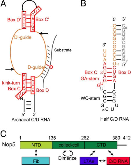
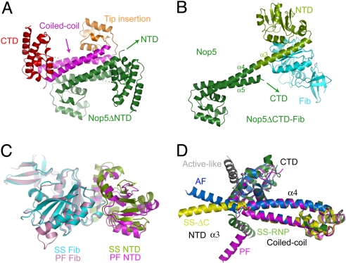
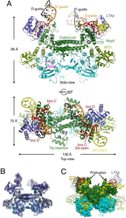
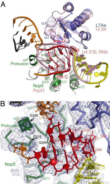
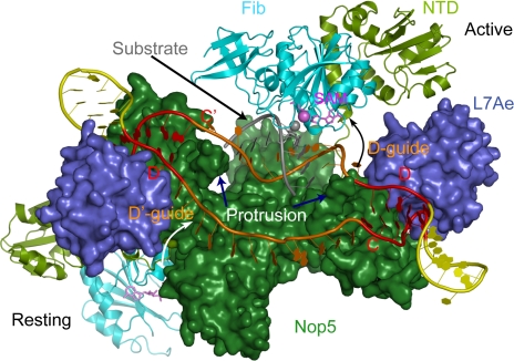
Box C/D guide RNAs are abundant noncoding RNAs that primarily function to direct the 2'-O-methylation of specific nucleotides by base-pairing with substrate RNAs. In archaea, a bipartite C/D RNA assembles with L7Ae, Nop5, and the methyltransferase fibrillarin into a modification enzyme with unique substrate specificity. Here, we determined the crystal structure of an archaeal C/D RNA-protein complex (RNP) composed of all 3 core proteins and an engineered half-guide RNA at 4 A resolution, as well as 2 protein substructures at higher resolution. The RNP structure reveals that the C-terminal domains of Nop5 in the dimeric complex provide symmetric anchoring sites for 2 L7Ae-associated kink-turn motifs of the C/D RNA. A prominent protrusion in Nop5 seems to be important for guide RNA organization and function and for discriminating the structurally related U4 snRNA. Multiple conformations of the N-terminal domain of Nop5 and its associated fibrillarin in different structures indicate the inherent flexibility of the catalytic module, suggesting that a swinging motion of the catalytic module is part of the enzyme mechanism. We also built a model of a native C/D RNP with substrate and fibrillarin in an active conformation. Our results provide insight into the overall organization and mechanism of action of C/D RNA-guided RNA methyltransferases.


Conflict of interest statement

The authors declare no conflict of interest.

Figures

Fig. 1.
Fig. 1.
Natural and half-C/D RNAs. (A) Schematic of the bipartite structure of an archaeal C/D RNA. The terminal boxes C and D form a canonical K-turn, whereas the internal boxes C′ and D′ fold into a K-loop. Boxes C/C′ and D/D′ are represented by their consensus sequences surrounded by red boxes. The guide upstream of box D (D-guide) is shown to associate with the substrate (gray), whereas the guide upstream of box D′ (D′-guide) is unbound. The hollow red circle strands for the modification target that is paired with the 5th nucleotide upstream of box D. Arrows indicate the cutting sites for generating the half-C/D RNA. (B) The sequence and secondary structure of the half-C/D RNA used in crystallization. The guide region pairs with the same sequence (gray hollow letters) from a crystallographic neighboring RNP. (C) Domain organization and interaction network of Nop5. The NTD, the coiled-coil domain, and the CTD are indicated. Numbers indicate the domain boundaries. Arrows indicate interaction relationships among RNP components.
Fig. 2.
Fig. 2.
The structures of Nop5 and fibrillarin. (A) The structure of dimeric Nop5ΔNTD. One subunit is colored deep blue, and the other is colored according to individual domains, with the CTD in red, the coiled-coil domain in magenta, and the tip insertion domain in orange. Dots strand for disordered loops. (B) Structure of the Nop5ΔCTD–Fib complex. Only a single copy is shown. The coiled-coil domain has the same orientation as in Fig. 2A. The CTD and NTD of Nop5 are colored dark and light green, respectively, and fibrillarin (Fib) is indicated in cyan. (C) The SS and PF Nop5 NTD–Fib complex structures aligned by fibrillarin. (D) Variable orientation between the NTD and the coiled-coil domain of Nop5. Nop5 structures in the SS Nop5ΔCTD–Fib (SS-ΔC, yellow), SS C/D RNP (SS-RNP, green), PF Nop5–Fib (PF, magenta), and AF Nop5–Fib (AF, blue) complexes, as well as an active model (active-like, gray), are shown. Only the α3 helix of the Nop5 NTD is shown for clarity. These structures are aligned on the basis of their CTDs and coiled-coil domains.
Fig. 3.
Fig. 3.
Overview of the C/D RNP structure. (A) Ribbon representation of the C/D RNP structure shown in side (Top) and top (Bottom) views. Boxes C, D, C′, and D′ are red, the guides are orange, the rest of the RNA is yellow, and the pairing guides from a crystallographic molecule are indicated in gray. The coiled-coil domain and CTD of Nop5 are dark green, the NTD of Nop5 is light green, L7Ae is blue, and fibrillarin is cyan. SAM is represented by magenta sticks. (B) Experimental electron density surrounding the entire C/D RNP structure. The map is contoured at 1.5σ, and the C/D RNP structure is shown as Ca or P traces in the side view. (C) Conserved surfaces in Nop5 and fibrillarin. Residues conserved in >90% of 56 archaeal Nop5 and fibrillarin sequences are colored yellow. None of the residues in the Nop5 NTD meet this conservation cutoff. The 2 copies of Nop5 and fibrillarin in the dimeric structure display nearly opposite surfaces. This view is slightly tilted from the side view to maximize visualization of the conserved region. The Nop5 NTD and L7Ae are shown as Cα traces. The guide regions of the RNA are not shown for clarity.
Fig. 4.
Fig. 4.
Recognition of the C/D RNA. (A) Structural superposition of the C/D RNP and the 15.5K–Prp31–U4 5′SL snRNP complex. The C/D RNP is color-coded as in Fig. 3A, and the U4 snRNP is pink. (B) Interaction between the Nop5 CTD and the half C/D RNA. The σA-weighted 2Fo-Fc map is shown at the 1.0σ contour level. RNAs and candidate residues for specific RNA recognition are shown as sticks, and proteins are shown as Cα traces. The protrusion boundaries (G299 and G318) are indicated by arrows.
Fig. 5.
Fig. 5.
Model of the native C/D RNP in resting and active states. The C/D half of the RNP complex is in the active state, with the D-guide forming a 9-bp duplex with the substrate and fibrillarin approaching the substrate. The 2′-OH group of the target nucleotide paired to the 5th nt upstream of box D is shown as a gray sphere, and the SAM methyl group to be transferred is shown as a violet sphere. The other half of the complex is in a resting state, with boxes C and D′ connected by a linear 12-nt guide. The curved arrows indicate the transition path for the catalytic module from the resting state to the active state.

References

    1. Czerwoniec A, et al. MODOMICS: A database of RNA modification pathways. 2008 update. Nucleic Acids Res. 2009;37:D118–D121. - PMC - PubMed
    1. Maxwell ES, Fournier MJ. The small nucleolar RNAs. Annu Rev Biochem. 1995;64:897–934. - PubMed
    1. Venema J, Tollervey D. Ribosome synthesis in Saccharomyces cerevisiae. Annu Rev Genet. 1999;33:261–311. - PubMed
    1. Kiss T. Small nucleolar RNA-guided post-transcriptional modification of cellular RNAs. EMBO J. 2001;20:3617–3622. - PMC - PubMed
    1. Omer AD, et al. RNA-modifying machines in archaea. Mol Microbiol. 2003;48:617–629. - PubMed

Publication types

LinkOut - more resources