. 2009 Aug 18;106(33):14022-7.
doi: 10.1073/pnas.0903201106. Epub 2009 Aug 5.

Allogeneic mesenchymal stem cells restore cardiac function in chronic ischemic cardiomyopathy via trilineage differentiating capacity

Affiliations

Allogeneic mesenchymal stem cells restore cardiac function in chronic ischemic cardiomyopathy via trilineage differentiating capacity

Henry C Quevedo et al. Proc Natl Acad Sci U S A. .

Abstract

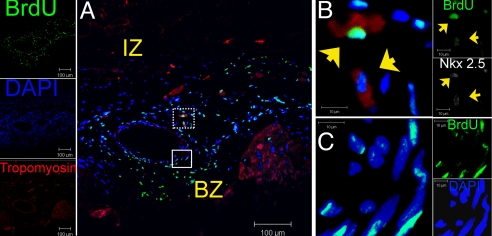
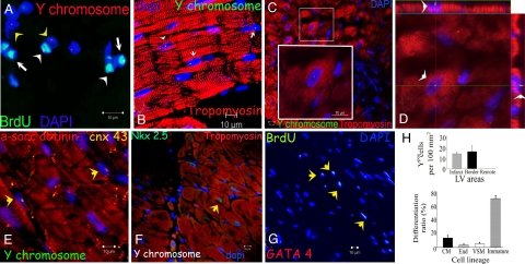
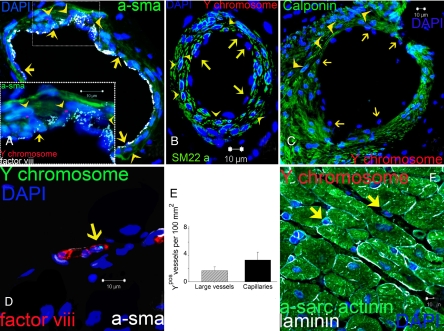
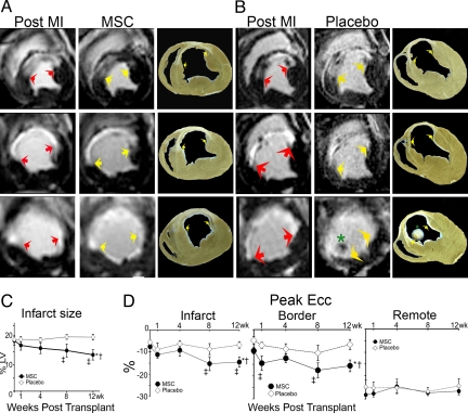
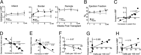
The mechanism(s) underlying cardiac reparative effects of bone marrow-derived mesenchymal stem cells (MSC) remain highly controversial. Here we tested the hypothesis that MSCs regenerate chronically infarcted myocardium through mechanisms comprising long-term engraftment and trilineage differentiation. Twelve weeks after myocardial infarction, female swine received catheter-based transendocardial injections of either placebo (n = 4) or male allogeneic MSCs (200 million; n = 6). Animals underwent serial cardiac magnetic resonance imaging, and in vivo cell fate was determined by co-localization of Y-chromosome (Y(pos)) cells with markers of cardiac, vascular muscle, and endothelial lineages. MSCs engrafted in infarct and border zones and differentiated into cardiomyocytes as ascertained by co-localization with GATA-4, Nkx2.5, and alpha-sarcomeric actin. In addition, Y(pos) MSCs exhibited vascular smooth muscle and endothelial cell differentiation, contributing to large and small vessel formation. Infarct size was reduced from 19.3 +/- 1.7% to 13.9 +/- 2.0% (P < 0.001), and ejection fraction (EF) increased from 35.0 +/- 1.7% to 41.3 +/- 2.7% (P < 0.05) in MSC but not placebo pigs over 12 weeks. This was accompanied by increases in regional contractility and myocardial blood flow (MBF), particularly in the infarct border zone. Importantly, MSC engraftment correlated with functional recovery in contractility (R = 0.85, P < 0.05) and MBF (R = 0.76, P < 0.01). Together these findings demonstrate long-term MSC survival, engraftment, and trilineage differentiation following transplantation into chronically scarred myocardium. MSCs are an adult stem cell with the capacity for cardiomyogenesis and vasculogenesis which contribute, at least in part, to their ability to repair chronically scarred myocardium.


Conflict of interest statement

The authors declare no conflict of interest.

Figures

Fig. 1.
Fig. 1.
Survival and distribution of transplanted MSCs in treated hearts. (A) Low power image of the infarct border region of MSC-treated heart depicting a band composing of BrdUpos cells (green) 12 weeks posttransplantation forming a medium-size vessel composed of BrdU pos cells throughout the vascular wall. (B) High magnification of the dotted square in (A) illustrating two BrdUpos nuclei (arrowheads) adjacent to the vessel, co-localized with cardiac transcription factor Nkx2.5 (white) and the structural protein tropomyosin (red) as depicted in the contiguous individual fluorescence channels on the right. (C) High power view of the square in (A) showing that BrdUpos cells are distributed throughout the vessel wall. Individual fluorescence channels for Nkx2.5, BrdU, and DAPI are shown in the adjacent insets. For double immunofluorescence co-localizations, n = 4 for both MSC and placebo hearts. At least four tissue sections from infarct, border, and remote zone were evaluated per heart. IZ, infarct zone; BZ, border zone.
Fig. 2.
Fig. 2.
Cardiogenic potential of transplanted MSCs. (A) Cluster of Ypos/BrdUpos cells (white arrowheads) located in infarct and border zones of treated hearts 12 weeks after MSCs implantation. Some of the transplanted MSCs do not exhibit BrdUpos signal (green), but maintain Ypos signal (red, yellow arrowheads). Conversely, another group shows BrdUpos signal (white arrows) and negative Y chromosome signal due technique sensitivity. (B) Cluster of Ypos cells (green, white arrows) in the border zone of MSC-treated animals co-localizing with tropomyosin (red). (C) Evidence of cardiac differentiation in a panoramic view of an infarct border zone of MSC-treated hearts. The inset depicts one Ypos (green) myocyte co-stained with tropomyosin. High magnification of the square is shown in the inset. (D) Confocal microscopy analysis of the same cell by orthogonal section of a z-stack (arrows point the cell analyzed in xy-plane). (E) Two transplanted Ypos cells (green, arrows) coupled with the resident cardiomyocytes by expressing connexin-43 (orange). (F) Evidence of cardiac commitment in the transplanted cell by the co-localization of Ypos signal with the cardiac transcription factor Nkx2.5 (green, arrow). Nuclei were counterstained with DAPI in all of the immunofluorescence assays. (G) Cluster of BrdUpos cells (green) in the border zone of MSC-treated animals exhibiting co-localization with transcription factor GATA-4 (red, arrows). (H) Quantitation of transplanted cells according to Y chromosome cell tracking. Ypos cells show no preference in distribution according to LV areas (top). Importantly, at 12 weeks posttransplantation, implanted MSCs showed commitment to repopulate the three major cardiac cell lineages and maintain a reservoir of nondifferentiated cells (bottom). Cell quantification per unit area for the Y chromosome (n = 6 for MSC-treated hearts, n = 4 for placebo-treated hearts). At least four tissue sections for infarct, border, and remote zone per heart were evaluated. Total area evaluated 2,673.34 mm2. CM, cardiomyocyte; End, endothelial cells; VSM, vascular smooth muscle.
Fig. 3.
Fig. 3.
Vascular differentiation of transplanted MSCs. (A) Representative image of a vessel containing numerous Ypos cells co-localized with smooth muscle actinin (a-sma in green, arrowheads) and endothelial cells (factor VIII-related antigen in white, arrows). High magnification of the inset to visualize the Ypos cells that co-localize with sma (arrowheads) and factor VIII-related antigen (white, arrows) demonstrating vascular smooth muscle and endothelial commitment, respectively. (B and C) Confirmation of Ypos cells commitment into vascular structures as depicted by co-localization with SM22-α (B) and calponin (C, arrowheads in both pictures). Ypos cells also commit to endothelial cell lineages (arrows). (D) Capillary formation with the incorporation of Ypos cell (arrow) co-stained with factor VIII-related antigen depicting the luminal surface of the vessel. (E) Assessment of vessel number per unit area according to their respectively size. (F) Ypos cells also reside in the interstitial compartment (arrows) of border myocardium in a nondifferentiated stage (n = 6 for MSC-treated hearts, n = 4 for placebo). At least 4 tissue sections from infarct, border, and remote zone were evaluated per animal.
Fig. 4.
Fig. 4.
Infarct size assessment and regional myocardial function. (A and B) Sequential short axis heart sections from base (top) to apex (bottom) of delayed gadolinium enhancement MRI images depicting the infarct extension (white) before treatment and 12 weeks following MSC therapy (A) compared with the placebo (B). Comparable gross pathology sections are shown adjacent to the MRI images. Arrows delineate the infarct extension, and the asterisk illustrates the presence of a thrombus near the apex in a placebo animal. (C) Reduction in infarct size following 8 and 12 weeks post MSC transplantation versus placebo (*P < 0.001 within MSC group ANOVA, P < 0.05 between groups ANOVA, P < 0.001 vs. preinjection status by Student-Newman-Keuls test). (D) Myocardial strain analysis represented by peak Ecc decreased in response to MSC-treatment in infarct (Left) and border (Center) areas but remained constant in the remote uninfarcted zone (Right) (*P < 0.05 for ANOVA within MSC group, P < 0.05 ANOVA between groups, P < 0.05 vs. preinjection status by Student-Newman-Keuls test). At least five MRI time points were analyzed for MSC-treated hearts (n = 6) and placebo-treated hearts (n = 4). For week-4 tagged MRI images and week-8 delayed contrast enhancement MRI analysis, MSCs (n = 4) and placebo (n = 4).
Fig. 5.
Fig. 5.
Myocardial blood flow and segmental contractility showed improvement related to the cell engraftment. (A) Quantitative analysis of first-pass perfusion MRI in the infarct (Left), border (Center), and remote (Right) myocardium demonstrating the improvement in the MSC-treated group compared to placebo group (*P < 0.05 for ANOVA within MSC, P < 0.001 ANOVA between groups, P < 0.05 vs. preinjection status by Student-Newman-Keuls test; LVBP, left ventricular blood pool). (B) LV ejection fraction improves in MSC group at 12 weeks posttransplantation (*P < 0.05 for ANOVA within MSC, P < 0.05 ANOVA between groups, P < 0.05 vs. preinjection status by Student-Newman-Keuls). (C–H) The functional outcomes in heart function (i.e., infarct size reduction, increase in contractility and increase in tissue perfusion) showed related interaction between them (C–E) and with the magnitude of cells detected (F–H) at 12 weeks after injection (P < 0.05 for all Pearson correlations). At least five time points for first-pass perfusion MRI image analysis. MSCs-treated hearts (n = 6) and placebo (n = 4) hearts for most of the time points except at 4 weeks for ejection fraction (n = 8, 4 MSC and 4 placebo).

Similar articles

Cited by

References

    1. Chien KR, Domian IJ, Parker KK. Cardiogenesis and the complex biology of regenerative cardiovascular medicine. Science. 2008;322:1494–1497. - PubMed
    1. Assmus B, et al. Transcoronary transplantation of progenitor cells after myocardial infarction. N Engl J Med. 2006;355:1222–1232. - PubMed
    1. Meyer GP, et al. Intracoronary bone marrow cell transfer after myocardial infarction: Eighteen months' follow-up data from the randomized, controlled BOOST (bone marrow transfer to enhance ST-elevation infarct regeneration) trial. Circulation. 2006;113:1287–1294. - PubMed
    1. Psaltis PJ, Zannettino AC, Worthley SG, Gronthos S. Concise review: Mesenchymal stromal cells: Potential for cardiovascular repair. Stem Cells. 2008;26:2201–2210. - PubMed
    1. Belema-Bedada F, Uchida S, Martire A, Kostin S, Braun T. Efficient homing of multipotent adult mesenchymal stem cells depends on FROUNT-mediated clustering of CCR2. Cell Stem Cell. 2008;2:566–575. - PubMed

Publication types