. 2009 Aug 11;106(32):13541-5.
doi: 10.1073/pnas.0906851106. Epub 2009 Jul 28.

Negative membrane curvature as a cue for subcellular localization of a bacterial protein

Affiliations

Negative membrane curvature as a cue for subcellular localization of a bacterial protein

Kumaran S Ramamurthi et al. Proc Natl Acad Sci U S A. .

Abstract

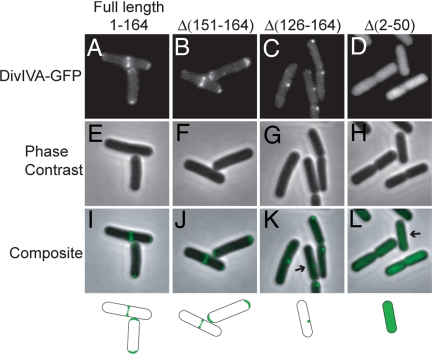
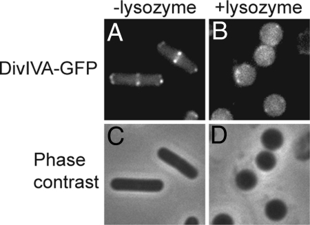
Bacterial proteins often localize to distinct sites within the cell, but the primary cues that dictate localization are largely unknown. Recent evidence has shown that positive membrane curvature can serve as a cue for localization of a peripheral membrane protein. Here we report that localization of the peripheral membrane protein DivIVA is determined in whole or in part by recognition of negative membrane curvature and that regions of the protein near the N and C terminus are important for localization. DivIVA, which is a cell division protein in Bacillus subtilis, localizes principally as a ring at nascent septa and secondarily to the less negatively curved, inside surface of the hemispherical poles of the cell. When cytokinesis is prevented, DivIVA redistributes itself to, and becomes markedly enriched at, the poles. When the rod-shaped cells are converted into spheres (protoplasts) by treatment with lysozyme, DivIVA adopts a largely uniform distribution around the cell. Recognition of membrane curvature by peripheral membrane proteins could be a general strategy for protein localization in bacteria.


Conflict of interest statement

The authors declare no conflict of interest.

Figures

Fig. 1.
Fig. 1.
Geometric cues for bacterial protein localization. (A) A cross-section of a sporulating B. subtilis cell is represented in which the spherical developing spore displays the only convex, or positively curved, surface (yellow) in the cytosol of the surrounding, rod-shaped cell (blue) which SpoVM recognizes as a localization cue. (B) Schematic cross section of an actively dividing B. subtilis cell, which harbors two areas of extreme concave, or negative, curvature (yellow) that recruits DivIVA. The most sharply concave region is formed along the edge where the division septum (center of the cell) meets the periphery of the cell. A second, less extremely concave site is the inside surface of the hemispherical cell poles.
Fig. 2.
Fig. 2.
DivIVA-GFP localizes to septa and poles. (A) Localization of full-length DivIVA-GFP (strain KR528), and truncated versions (B) DivIVAΔ(151–164)-GFP (strain KR534), (C) DivIVAΔ(126–164)-GFP (strain KR533), and (D) DivIVAΔ(2–50)-GFP (strain KR536) in a sinI mutant in which cell chaining is prevented. Fusions were expressed from the native divIVA promoter. A–D and E–H are, respectively, fluorescence and phase contrast images of the same cells in each column. I–L are overlays of the corresponding fluorescence and phase contrast images.
Fig. 3.
Fig. 3.
DivIVA-GFP localizes to septa in anucleate cells. A and B show, respectively, localization of DivIVA-GFP and DivIVAΔ(2–50)-GFP in the wild type (strains KR541 and KR571). C–E show localization of DivIVA-GFP (C and D) and DivIVAΔ(2–50)-GFP (E) in cells overexpressing the DNA replication inhibition gene sirA (strains KR570 and KR572). D shows an example of adjacent anucleate cells that arise from SirA overproduction. A–E are composite images showing GFP fluorescence (green) and DNA (blue) overlaid on membrane (red). F–J show fluorescence from GFP, K–O fluorescence from membrane staining with the dye FM4–64, and P–T DNA staining with DAPI. gfp fusions and sirA was expressed from the inducible promoter hyperspank by the addition of IPTG. The same cells are shown from top to bottom in each column.
Fig. 4.
Fig. 4.
DivIVA-GFP redistributes from septa to poles upon inhibition of cytokinesis. (A) DivIVA-GFP localization in wild type strain (KR568). (B–D) DivIVA-GFP localization in cells overexpressing mciZ under the control of xylose-inducible promoter at the indicated times after addition of xylose (strain KR569). (E–H) Membranes of cells in A–D visualized with the dye FM4–64. GFP fusions were expressed from the IPTG-inducible hyperspank promoter.
Fig. 5.
Fig. 5.
DivIVA-GFP localizes uniformly in protoplasts. (A) DivIVA-GFP localization in sinI mutant cells that were not treated with lysozyme or (B) that were treated with lysozyme. (C and D) Phase contrast images of cells in A and B.

References

    1. Blobel G. Intracellular protein topogenesis. Proc Natl Acad Sci USA. 1980;77:1496–1500. - PMC - PubMed
    1. Shapiro L, McAdams HH, Losick R. Generating and exploiting polarity in bacteria. Science. 2002;298:1942–1946. - PubMed
    1. Ramamurthi KS, Lecuyer S, Stone HA, Losick R. Geometric cue for protein localization in a bacterium. Science. 2009;323:1354–1357. - PMC - PubMed
    1. Stragier P, Losick R. Molecular genetics of sporulation in Bacillus subtilis. Annu Rev Genet. 1996;30:297–241. - PubMed
    1. Kim H, et al. The Bacillus subtilis spore coat protein interaction network. Mol Microbiol. 2006;59:487–502. - PubMed

Publication types

MeSH terms

LinkOut - more resources