. 2009 Aug 18;106(33):13820-5.
doi: 10.1073/pnas.0905718106. Epub 2009 Aug 3.

Residual breast cancers after conventional therapy display mesenchymal as well as tumor-initiating features

Affiliations

Residual breast cancers after conventional therapy display mesenchymal as well as tumor-initiating features

Chad J Creighton et al. Proc Natl Acad Sci U S A. .

Abstract

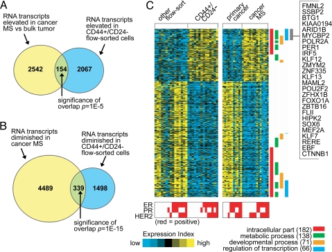
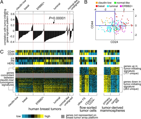
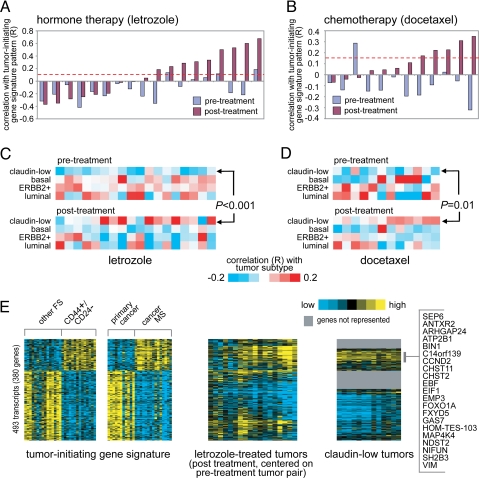
Some breast cancers have been shown to contain a small fraction of cells characterized by CD44(+)/CD24(-/low) cell-surface antigen profile that have high tumor-initiating potential. In addition, breast cancer cells propagated in vitro as mammospheres (MSs) have also been shown to be enriched for cells capable of self-renewal. In this study, we have defined a gene expression signature common to both CD44(+)/CD24(-/low) and MS-forming cells. To examine its clinical significance, we determined whether tumor cells surviving after conventional treatments were enriched for cells bearing this CD44(+)/CD24(-/low)-MS signature. The CD44(+)/CD24(-/low)-MS signature was found mainly in human breast tumors of the recently identified "claudin-low" molecular subtype, which is characterized by expression of many epithelial-mesenchymal-transition (EMT)-associated genes. Both CD44(+)/CD24(-/low)-MS and claudin-low signatures were more pronounced in tumor tissue remaining after either endocrine therapy (letrozole) or chemotherapy (docetaxel), consistent with the selective survival of tumor-initiating cells posttreatment. We confirmed an increased expression of mesenchymal markers, including vimentin (VIM) in cytokeratin-positive epithelial cells metalloproteinase 2 (MMP2), in two separate sets of postletrozole vs. pretreatment specimens. Taken together, these data provide supporting evidence that the residual breast tumor cell populations surviving after conventional treatment may be enriched for subpopulations of cells with both tumor-initiating and mesenchymal features. Targeting proteins involved in EMT may provide a therapeutic strategy for eliminating surviving cells to prevent recurrence and improve long-term survival in breast cancer patients.


Conflict of interest statement

The authors declare no conflict of interest.

Figures

Fig. 1.
Fig. 1.
A gene transcription signature of breast cancer cells with putative tumor-initiating potential. (A) Venn diagram of the intersection between genes elevated in cancer mammospheres (MSs, rich in CD44+/CD24−/low cells) compared with primary cancers and genes elevated in CD44+/CD24−/low cells obtained from FACS assay (P < 0.01, fold change >1.5 for each comparison). P value for the overlap between the two gene sets by one-sided Fisher's exact test. (B) As with A but for genes lower in cancer MSs and genes lower in CD44+/CD24−/low flow-sorted cells. (C) Heat map of genes in the CD44+/CD24−/low-MS signature (from parts A and B). Each row represents a transcript; each column, a sample (yellow: high expression). Associations of the genes with selected Gene Ontology (GO) annotation terms are indicated. Genes in the indicated region are listed by name.
Fig. 2.
Fig. 2.
The CD44+/CD24−/low-MS gene signature is enriched in human breast tumors of the claudin-low molecular subtype. (A) Correlation (R value, see Methods) between the CD44+/CD24−/low-MS signature pattern and each tumor in the gene expression profile dataset by Herschkowitz et al. (6) (claudin-low, basal, ERBB2+, luminal, normal-like; averages centered on the mean centroid of the groups). R values above red dotted line are significant at P < 0.00001. (B) Scatter plot of CD24 versus CD44 mRNA expression for the Herschkowitz tumors. (C) Heat map of the corresponding patterns of the CD44+/CD24−/low-MS signature genes within the Herschkowitz tumors. The order of the Herschkowitz tumors is the same for A and C; the order of the genes is the same across datasets.
Fig. 3.
Fig. 3.
The CD44+/CD24−/low-MS gene signature is enriched in a subset of human breast tumors after treatment with hormone therapy or chemotherapy. (A) Correlation between the CD44+/CD24−/low-MS signature pattern and each of 36 human breast tumor profiles, representing 18 pairs before and after treatment with letrozole. R values above red dotted line are significant at P < 0.01. (B) Correlation between the CD44+/CD24−/low-MS signature pattern and each of 24 tumor profiles, representing 12 pairs before and after treatment with docetaxel. (C) Heat map of the correlations between each letrozole-treated tumor profile (part A) and the average expression for each of the four major Herschkowitz molecular tumor profile subtypes (claudin-low, basal, ERBB2+, luminal; averages centered on the mean centroid of the groups). P value comparing pretreatment and posttreatment R values for the claudin-low group by paired t test. (D) As with part C, but for the docetaxel-treated tumors (B). (E) Heat maps of the corresponding patterns of the CD44+/CD24−/low-MS signature genes within the letrozole posttreatment tumors (each tumor centered on corresponding pretreatment pair) and the Herschkowitz claudin-low tumors (centered on mean centroid of tumor groups). Gene ordering is the same across datasets.
Fig. 4.
Fig. 4.
Specific markers of mesenchymal cells are overexpressed in both CD44+/CD24−/low-MS cells and letrozole-treated patient tumors. (A) For the flow sort and cancer MS profile datasets, heat map of previously associated mesenchymal genes in a review by Lee et al. (31) (genes in bold, significant with P < 0.01; the entire set of mesenchymal-associated proteins from the review that we could map to specific genes in our dataset are represented). (B) Protein expression of vimentin and pan-cytokeratin (CK) in a patient tumor after letrozole treatment. (C) Immunofluorescence analysis of vimentin (red) and pan-cytokeratin (green) after chemotherapy. Yellow arrows mark double-positive cells, whereas red and green arrows mark vimentin and CK positive epithelial cells, respectively. (D) MMP2mRNA levels in tumors before and after letrozole. (P values in B and C by paired t test).

Comment in

  • The changing faces of cancer cells.
    Feng XH. Feng XH. Nat Rev Mol Cell Biol. 2010 Jul;11(7):466. doi: 10.1038/nrm2923. Epub 2010 Jun 16. Nat Rev Mol Cell Biol. 2010. PMID: 20551963 No abstract available.

Similar articles

Cited by

References

    1. Dick JE. Stem cell concepts renew cancer research. Blood. 2008;112:4793–4807. - PubMed
    1. Al-Hajj M, Wicha MS, Benito-Hernandez A, Morrison SJ, Clarke MF. Prospective identification of tumorigenic breast cancer cells. Proc Natl Acad Sci USA. 2003;100:3983–3988. - PMC - PubMed
    1. Li X, et al. Intrinsic resistance of tumorigenic breast cancer cells to chemotherapy. J Natl Cancer Inst. 2008;100:672–679. - PubMed
    1. Sorlie T, et al. Gene expression patterns of breast carcinomas distinguish tumor subclasses with clinical implications. Proc Natl Acad Sci USA. 2001;98:10869–10874. - PMC - PubMed
    1. Perou CM, et al. Molecular portraits of human breast tumours. Nature. 2000;406:747–752. - PubMed

Publication types