

Conservation of enhancer location in divergent insects
- PMID: 19666595
- PMCID: PMC2732830
- DOI: 10.1073/pnas.0905754106
Conservation of enhancer location in divergent insects
Abstract
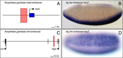
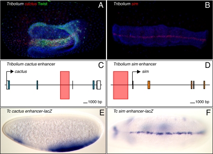
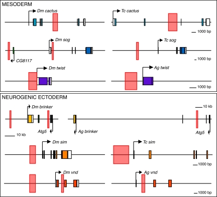
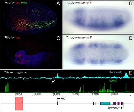
Dorsoventral (DV) patterning of the Drosophila embryo is controlled by a concentration gradient of Dorsal, a sequence-specific transcription factor related to mammalian NF-kappaB. The Dorsal gradient generates at least 3 distinct thresholds of gene activity and tissue specification by the differential regulation of target enhancers containing distinctive combinations of binding sites for Dorsal, Twist, Snail, and other DV determinants. To understand the evolution of DV patterning mechanisms, we identified and characterized Dorsal target enhancers from the mosquito Anopheles gambiae and the flour beetle Tribolium castaneum. Putative orthologous enhancers are located in similar positions relative to the target genes they control, even though they lack sequence conservation and sometimes produce divergent patterns of gene expression. The most dramatic example of this conservation is seen for the "shadow" enhancer regulating brinker: It is conserved within the intron of the neighboring Atg5 locus of both flies and mosquitoes. These results suggest that, like exons, an enhancer position might be subject to constraint. Thus, novel patterns of gene expression might arise from the modification of conserved enhancers rather than the invention of new ones. We propose that this enhancer constancy might be a general property of regulatory evolution, and should facilitate enhancer discovery in nonmodel organisms.
Conflict of interest statement
The authors declare no conflict of interest.
Figures
References
-
- Stathopoulos A, Van Drenth M, Erives A, Markstein M, Levine M. Whole-genome analysis of dorsal-ventral patterning in the Drosophila embryo. Cell. 2002;111:687–701. - PubMed
-
- Stein D, Nusslein-Volhard C. Multiple extracellular activities in Drosophila egg perivitelline fluid are required for establishment of embryonic dorsal-ventral polarity. Cell. 1992;68:429–440. - PubMed
-
- Roth S, Hiromi Y, Godt D, Nusslein-Volhard C. cactus, a maternal gene required for proper formation of the dorsoventral morphogen gradient in Drosophila embryos. Development. 1991;112:371–388. - PubMed
-
- Moussian B, Roth S. Dorsoventral axis formation in the Drosophila embryo-shaping and transducing a morphogen gradient. Curr Biol. 2005;15:R887–R899. - PubMed
-
- Stathopoulos A, Levine M. Dorsal gradient networks in the Drosophila embryo. Dev Biol. 2002;246:57–67. - PubMed
Publication types
MeSH terms
Substances
Grants and funding
LinkOut - more resources
Full Text Sources
Molecular Biology Databases
Research Materials
