. 2009 Aug 18;106(33):13826-31.
doi: 10.1073/pnas.0901259106. Epub 2009 Aug 3.

Human BAHD1 promotes heterochromatic gene silencing

Affiliations

Human BAHD1 promotes heterochromatic gene silencing

Hélène Bierne et al. Proc Natl Acad Sci U S A. .

Abstract

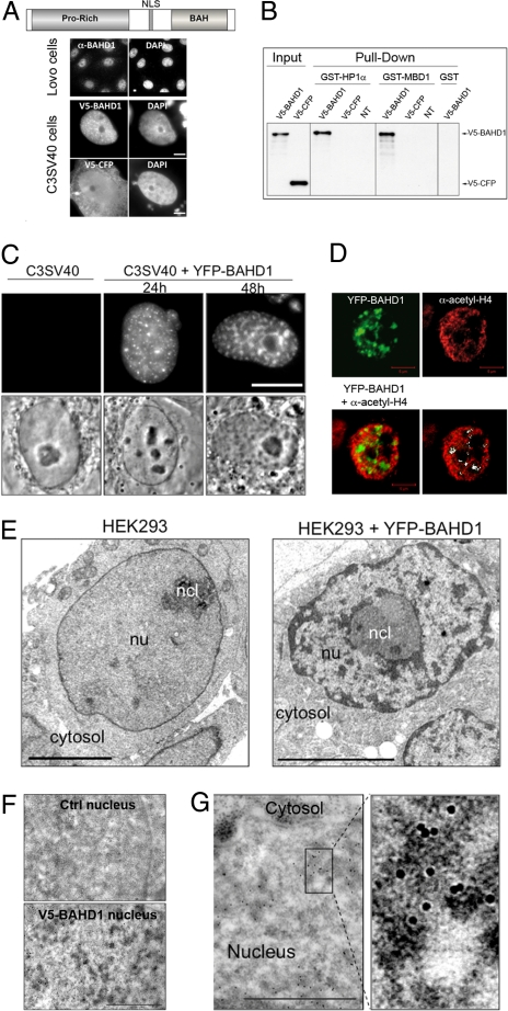
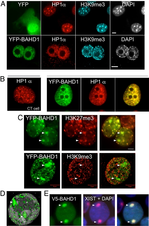
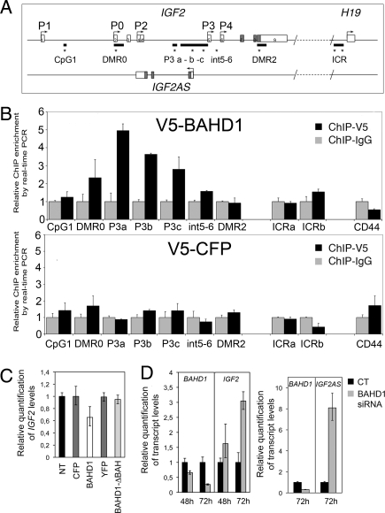
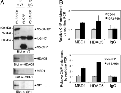
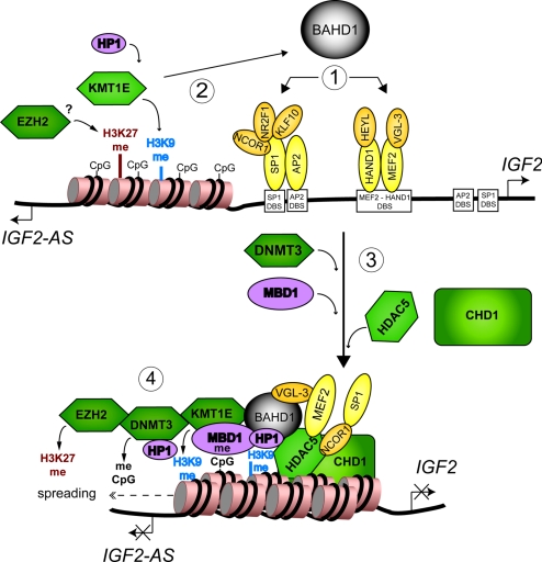
Gene silencing via heterochromatin formation plays a major role in cell differentiation and maintenance of homeostasis. Here we report the identification and characterization of a novel heterochromatinization factor in vertebrates, bromo adjacent homology domain-containing protein 1 (BAHD1). This nuclear protein interacts with HP1, MBD1, HDAC5, and several transcription factors. Through electron and immunofluorescence microscopy studies, we show that BAHD1 overexpression directs HP1 to specific nuclear sites and promotes the formation of large heterochromatic domains, which lack acetyl histone H4 and are enriched in H3 trimethylated at lysine 27 (H3K27me3). Furthermore, ectopically expressed BAHD1 colocalizes with the heterochromatic inactive X chromosome (Xi). The BAH domain is required for BAHD1 colocalization with H3K27me3, but not with the Xi chromosome. As highlighted by whole genome microarray analysis of BAHD1 knockdown cells, BAHD1 represses several proliferation and survival genes, in particular the insulin-like growth factor II gene (IGF2). When overexpressed, BAHD1 specifically binds the CpG-rich P3 promoter of IGF2, which increases MBD1 and HDAC5 targeting at this locus. This region contains DNA-binding sequences for the transcription factor SP1, with which BAHD1 coimmunoprecipitates. Collectively, these findings provide evidence that BAHD1 acts as a silencer by recruiting at specific promoters a set of proteins that coordinate heterochromatin assembly.


Conflict of interest statement

The authors declare no conflict of interest.

Figures

Fig. 1.
Fig. 1.
BAHD1 promotes heterochromatin formation. (A) Endogenous BAHD1 localizes predominantly to nuclei, labeled with DAPI, when detected with an anti-BAHD1 antibody in Lovo epithelial cells. V5-BAHD1 expressed in C3SV40 fibroblasts localizes only to the nucleus, whereas control V5-CFP localizes to both the cytosol and the nucleus. (Scale bar: 5 μm.) (B) BAHD1 binds HP1α and MBD1. Nuclear extracts from HEK293 cells either untransfected (NT) or transfected with V5-BAHD1 or V5-CFP constructs were incubated with purified GST-HP1α, GST-MBD1, or GST. Bound BAHD1 was detected in immunoblots using anti-V5 antibody; 10% of nuclear extracts were analyzed in parallel (input). (C–E) Increasing the amount of BAHD1 in cells stimulates chromatin condensation. (C) In living C3SV40 cells, YFP-BAHD1 forms nuclear foci that increase in size with increasing transfection time (24–48 h) and appear as dense material in phase contrast (Movie S1). Large dark bodies are nucleoli. (Scale bar: 10 μm.) (D) Confocal image of a YFP-BAHD1–transfected C3SV40 cell labeled with acetyl-H4 antibody. The white regions on the last image are positive for YFP-BAHD1 and negative for acetyl-H4. (Scale bar: 5 μm.) (E) Transmission electron microscopy reveals dense patches of condensed chromatin formed in nucleus of YFP-BAHD1 expressing HEK293 cells. (Scale bar: 5 μm.) (F) Anti-V5 immunogold cytochemistry demonstrates localization of V5-BAHD1 at de novo formed heterochromatin. V5-BAHD1, detected with 10-nm colloidal gold particles, specifically localizes to electron-dense areas in the nucleus that are not present in untransfected cells (Ctrl). (Scale bar: 1 μm.) (G) High magnification highlights localization of V5-BAHD particles within electron-dense heterochromatin areas.
Fig. 2.
Fig. 2.
BAHD1 recruits HP1α and colocalizes with H3K27me3 in vivo. (A) BAHD1 delocalizes HP1α from chromocenters in mouse fibroblasts. In 3T3 fibroblasts expressing YFP, HP1α localizes to chromocenter nuclear bodies labeled with H3K9me3 antibodies and DAPI. In YFP-BAHD1–transfected cells, HP1α no longer accumulates at chromocenters, but instead localizes mainly at YFP-BAHD1 discrete foci (Fig. S2). (Scale bar: 5 μm.) (B) HP1α is recruited to BAHD1-associated heterochromatin in human fibroblasts. In C3SV40 cells expressing control YFP (CT cell), HP1α localizes at dispersed nuclear foci. In YFP-BAHD1–expressing cells, HP1α colocalizes with BAHD1, as shown by yellow regions in the merge image. (C and D) YFP-BAHD1 heterochromatic foci have features of facultative heterochromatin. (C) YFP-BAHD1 expressed in HEK293 cells colocalizes with H3K27me3 at several nuclear territories, including Xi (indicated by triangles). YFP-BAHD1 does not colocalize with H3K9me3. (Scale bar: 5 μm.) (D) Three-dimensional reconstruction of confocal images shows localization of V5-BAHD1 at the 2 Xi chromosomes (indicated by triangles), as well as several other domains dispersed in a HEK293 cell nucleus (Movie S2). (E) Exogenous V5-BAHD1 is recruited to the Xi chromosomes when expressed for 24 h in HEK293 cells. The Xi chromosomes were detected by RNA FISH using a Xist probe (in red) in the nucleus labeled with DAPI (in blue).
Fig. 3.
Fig. 3.
BAHD1 targets IGF2. (A) Organization of the human IGF2/IGF2AS/H19 genomic region. Shown are the positions of IGF2 promoters (P0→P4), CpG islands (black lines), exons (open boxes), and ORF (gray boxes) (adapted from ref. 26). Stars indicate the position of primers used in ChIP. The position of IGF2AS on the opposite strand is shown. (B) BAHD1 binds IGF2 in the P3 proximal region. ChIP assays with V5 antibodies or control IgG using chromatin prepared from V5-BAHD1– or control V5-CFP–expressing HEK293 cells. DNA fragments were quantified by qPCR with the primer sets indicated in (A). The CD44 promoter was used as a negative control because it shares features with IGF2 P3 (i.e., a CpG island and localization on chromosome 11). The amount of DNA precipitated with V5 antibody was normalized to the amount precipitated with IgG control. The data are averages (± SD) of qPCR replicates of 1 of 3 independent ChIP experiments. (C and D) BAHD1 represses IGF2 and IGF2AS. (C) HEK293 cells were transfected for 24 h with V5-CFP, V5-BAHD1, YFP, or BAHD1-BAH. The levels of IGF2 transcript quantified by qRT-PCR decreased with BAHD1 overexpression, but not with BAHD1-BAH overexpression. (D) HEK293 cells were transfected with BAHD1 siRNA or control siRNA (CT). BAHD1, IGF2, or IGF2AS transcript levels were quantified by qRT-PCR after 48 h or 72 h.
Fig. 4.
Fig. 4.
(A) Exogenous BAHD1 coimmunoprecipitates with endogenous HDAC5, MBD1, and SP1. Nuclear extracts from V5-BAHD1– or V5-CFP–expressing HEK293 cells were analyzed after IP with anti-V5 mAbs (IP α-V5) or with mouse IgG (IP IgG). Western blots were probed with antibodies against V5, HDAC5, MBD1, or SP1. IgG HC, heavy chains of immunoglobulins. (B) MBD1 and HDAC5 are recruited at IGF2 P3 in BAHD1-overexpressing cells. Chromatin from V5-BAHD1– or V5-CFP–expressing HEK293 cells was used in ChIP with MBD1 or HDAC5 antibodies or control IgG. P3b and CD44 DNA fragments were quantified by qPCR, as in Fig. 3B. The amount of DNA precipitated with the different antibodies was normalized to the amount precipitated with IgG. In cells expressing V5-BAHD1, MBD1 and HDAC5 recruitment at IGF2 P3b increased compared with that at the CD44 promoter (top) and was greater than in control cells expressing V5-CFP (bottom).
Fig. 5.
Fig. 5.
Hypothetical model for BAHD1-mediated heterochromatic silencing at the IGF2 P3 region. 1, BAHD1 targets IGF2 P3 by interacting with cofactors of DNA-bound transcription factors. 2, BAHD1 recruits KMT1E, which methylates K9 of histone H3 (H3K9me). HP1 is tethered to chromatin by binding H3K9me and BAHD1. EZH2 is recruited at this locus, before or after BAHD1, by an unknown mechanism and methylates K27 of H3 (H3K27me). 3, KMT1E and HP1 interact with DNMT3, which methylates cytosines (meCpG), creating binding sites for MBD1. BAHD1 recruits HDAC5 and CHD1, both of which bind NCOR1, which interacts with SP1. HDAC5 also binds MEF2. The complex is stabilized by cross-interactions between BAHD1, MBD1 and HDAC5 (Fig. S5). HDAC5 and CHD1 activities lead to histone deacetylation and nucleosome compaction. 4, Heterochromatin spreads to surrounding sequences after successive loading of BAHD1, histone, and DNA methylation marks, preventing access of the transcription machinery to the promoter and resulting in transcriptional repression of IGF2 and IGF2AS. Abnormal down-regulation of BAHD1 would reactivate IGF2 expression.

References

    1. Trojer P, Reinberg D. Facultative heterochromatin: Is there a distinctive molecular signature? Mol Cell. 2007;28:1–13. - PubMed
    1. Moss TJ, Wallrath LL. Connections between epigenetic gene silencing and human disease. Mutat Res. 2007;618:163–174. - PMC - PubMed
    1. Feinberg AP. Phenotypic plasticity and the epigenetics of human disease. Nature. 2007;447:433–440. - PubMed
    1. Wang GG, Allis CD, Chi P. Chromatin remodeling and cancer, part II: ATP-dependent chromatin remodeling. Trends Mol Med. 2007;13:373–380. - PMC - PubMed
    1. Wang GG, Allis CD, Chi P. Chromatin remodeling and cancer, part I: Covalent histone modifications. Trends Mol Med. 2007;13:363–372. - PubMed

Publication types

Associated data