. 2010 Jan 1;85(1):155-66.
doi: 10.1093/cvr/cvp275.

Endogenous generation and protective effects of nitro-fatty acids in a murine model of focal cardiac ischaemia and reperfusion

Affiliations

Endogenous generation and protective effects of nitro-fatty acids in a murine model of focal cardiac ischaemia and reperfusion

Volker Rudolph et al. Cardiovasc Res. .

Abstract

Aims: Nitrated fatty acids (NO(2)-FA) have been identified as endogenous anti-inflammatory signalling mediators generated by oxidative inflammatory reactions. Herein the in vivo generation of nitro-oleic acid (OA-NO(2)) and nitro-linoleic acid (LNO(2)) was measured in a murine model of myocardial ischaemia and reperfusion (I/R) and the effect of exogenous administration of OA-NO(2) on I/R injury was evaluated.

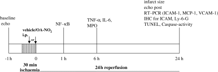
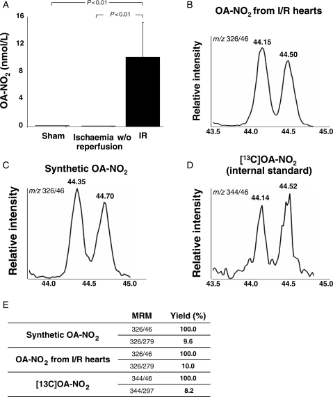
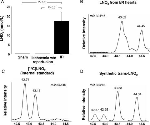
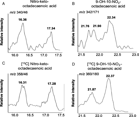
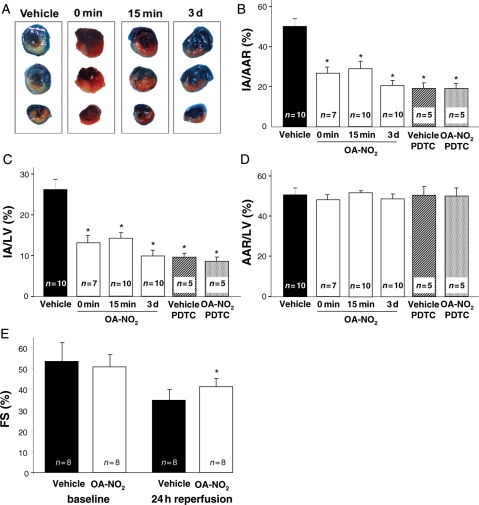
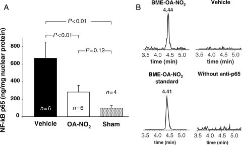
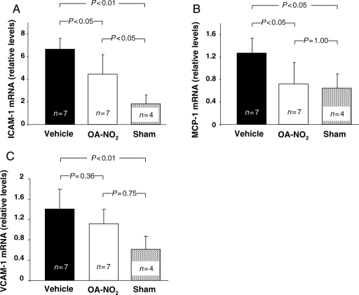
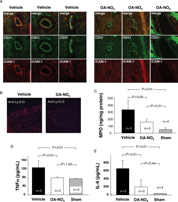
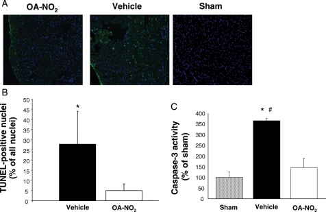
Methods and results: In C57/BL6 mice subjected to 30 min of coronary artery ligation, endogenous OA-NO(2) and LNO(2) formation was observed after 30 min of reperfusion, whereas no NO(2)-FA were detected in sham-operated mice and mice with myocardial infarction without reperfusion. Exogenous administration of 20 nmol/g body weight OA-NO(2) during the ischaemic episode induced profound protection against I/R injury with a 46% reduction in infarct size (normalized to area at risk) and a marked preservation of left ventricular function as assessed by transthoracic echocardiography, compared with vehicle-treated mice. Administration of OA-NO(2) inhibited activation of the p65 subunit of nuclear factor kappaB (NFkappaB) in I/R tissue. Experiments using the NFkappaB inhibitor pyrrolidinedithiocarbamate also support that protection lent by OA-NO(2) was in part mediated by inhibition of NFkappaB. OA-NO(2) inhibition of NFkappaB activation was accompanied by suppression of downstream intercellular adhesion molecule 1 and monocyte chemotactic protein 1 expression, neutrophil infiltration, and myocyte apoptosis.

Conclusion: This study reveals the de novo generation of fatty acid nitration products in vivo and reveals the anti-inflammatory and potential therapeutic actions of OA-NO(2) in myocardial I/R injury.


Figures

Scheme 1
Scheme 1
Experimental protocol for the assessment of OA-NO2 effects in the murine I/R model.
Figure 1
Figure 1
Endogenous generation of OA-NO2 following ischaemia and reperfusion. (A) Tissue concentration of OA-NO2 in hearts from mice undergoing 30 min of ischaemia and 30 min of reperfusion, ischaemia without reperfusion or sham surgery. (BD) Characterization of OA-NO2 in heart tissue from mice subjected to I/R injury. The chromatograms show co-elution of OA-NO2 extracted from heart tissue with synthetic OA-NO2 (C) and the internal standard (D). (E) The table shows the peak area ratios corresponding to the two main products of OA-NO2 collision induced dissociation: m/z 326/279 (neutral loss of HNO2) and m/z 326/46 (formation of NO2), further confirming the identity of OA-NO2 extracted from the heart tissue.
Figure 2
Figure 2
Endogenous generation of LNO2 following ischaemia and reperfusion. (A) Tissue concentration of LNO2 after I/R, ischaemia without reperfusion or sham surgery. (B) Chromatogram of LNO2 extracted from mouse hearts subjected to ischaemia and reperfusion. LNO2 eluted later than the [13C]-labelled cis-LNO2 which was used as internal standard (C) and co-eluted with synthetic trans-LNO2 (D). The slight shift in retention times between chromatograms (B and D) is due to variations between separate chromatographic runs.
Figure 3
Figure 3
LNO2 is further oxidized to keto (A) and hydroxyl (B) derivatives in murine heart tissue after I/R; and 3 µM [13C]LNO2 subjected to air oxidation overnight in Tris buffer (pH 8) was used as the precursor for oxidized 13C-labelled LNO2 derivatives. These species served as internal standards for identification of endogenously produced oxidized derivatives of LNO2 (C and D).
Figure 4
Figure 4
Effect of OA-NO2 on infarct size and left ventricular function following ischaemia and reperfusion. (A) Representative images of hearts from vehicle- or OA-NO2-treated animals stained with Evan's blue dye. (B) OA-NO2 treatment at reperfusion, 15 min or 3 days prior to reperfusion lead to a significant reduction in infarct area/area at risk ratio when compared with vehicle-treated animals (*P < 0.01 vs. vehicle). Pretreatment with pyrrolidinedithiocarbamate (PDTC) abolished the effect of OA-NO2. (C) Infarct area to left ventricular (IA/LV) areas reflected the results observed for the IA/AAR ratio (*P < 0.01 vs. vehicle). (D) No difference in AAR/LV ratio was observed between the different treatment groups. (E) OA-NO2 treatment resulted in a significantly improved left ventricular function after I/R, as assessed by transthoracic M-mode echocardiography (*P < 0.01).
Figure 5
Figure 5
Inhibition of NFκB activation by OA-NO2. (A) Reduction of NFκB p65 activity in myocardial tissue of OA-NO2 treated animals following I/R. (B) Assessment of nitroalkylation of p65 in myocardial tissue.
Figure 6
Figure 6
Effect of OA-NO2 on mRNA levels of adhesion molecules and MCP-1. (A) Reduced NFκB activation in OA-NO2-treated animals was accompanied by a significant reduction of ICAM-1 mRNA expression. (B) Significant suppression of MCP-1 mRNA levels in OA-NO2-treated animals. (C) VCAM-1 mRNA levels also showed a trend towards a reduction. All values are normalized to β2-microglobulin.
Figure 7
Figure 7
Effect of OA-NO2 on leukocyte infiltration and systemic inflammation following ischaemia and reperfusion. (A) Immunofluorescent staining for ICAM-1 (red) and CD31 (green) in vessels of OA-NO2 (right panel) and vehicle-treated animals (left panel). OA-NO2-treated animals showed a marked reduction in ICAM-1 immunoreactivity. (B) Neutrophil accumulation in infarcted zone of myocardium. Immunofluorescent staining for neutrophil-specific Ly-6-G revealed increased neutrophil infiltration in vehicle-treated mice. (C) Determination of MPO tissue levels confirmed increased neutrophil infiltration in vehicle-treated animals. (D) Serum TNFα levels were significantly reduced in OA-NO2 treated animals 6 h after reperfusion. (E) Serum IL-6 levels underwent significant reduction in OA-NO2-treated mice.
Figure 8
Figure 8
OA-NO2 reduced apoptosis in myocardial tissue after ischaemia and reperfusion. (A) Immunofluorescent staining for TUNEL-positive cells. (B) Quantification of TUNEL-positive cells; *P < 0.001. (C) Assessment of caspase 3 activity. *P < 0.05 for comparison with sham; #P < 0.05 for comparison with OA-NO2. No statistically significant difference was observed between OA-NO2 treated animals and sham animals.

References

    1. Yellon DM, Hausenloy DJ. Myocardial reperfusion injury. N Engl J Med. 2007;357:1121–1135. - PubMed
    1. Zweier JL, Talukder MA. The role of oxidants and free radicals in reperfusion injury. Cardiovasc Res. 2006;70:181–190. - PubMed
    1. Hearse DJ, Humphrey SM, Chain EB. Abrupt reoxygenation of the anoxic potassium-arrested perfused rat heart: a study of myocardial enzyme release. J Mol Cell Cardiol. 1973;5:395–407. - PubMed
    1. Granger DN, Rutili G, McCord JM. Superoxide radicals in feline intestinal ischemia. Gastroenterology. 1981;81:22–29. - PubMed
    1. Burton KP, McCord JM, Ghai G. Myocardial alterations due to free-radical generation. Am J Physiol. 1984;246:H776–H783. - PubMed

Publication types